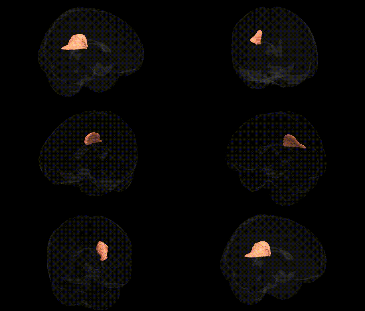
The bilateral Posterior corona radiata L, as defined in the JHU ICBM labels 1mm Atlas, refers to the left-sided portion of a major white matter tract within the corona radiata, located superior and posterior to the internal capsule and extending toward the parietal and occipital lobes. It contains predominantly projection fibers that connect the cerebral cortex with subcortical structures, including the thalamus and brainstem, and carries components of sensory, motor, and associative pathways important for integration of somatosensory information, visuospatial processing, and higher-level cognitive functions. As a part of the broader corona radiata, it is frequently analyzed in diffusion tensor imaging and tractography studies to assess white matter integrity in neurological and psychiatric conditions. There is no direct link for the Posterior corona radiata L; see the related structure Corona radiata (brain).
Genetic associations with the bilateral posterior corona radiata, as defined in the JHU ICBM labels 1 mm atlas, are inferred largely from GWAS of white matter microstructure and related neuropsychiatric traits rather than from region-specific candidate studies. Large diffusion MRI GWAS (e.g., ENIGMA-DTI and UK Biobank) have identified multiple loci influencing fractional anisotropy and mean diffusivity in posterior corona radiata tracts, implicating genes involved in axonal growth, myelination, and cytoskeletal organization (such as those near CNTN4, NTRK1/2, and other axon guidance or oligodendrocyte-related genes), though effects are typically polygenic and shared across multiple white matter bundles. Genetic variants associated with schizophrenia, bipolar disorder, major depression, and autism spectrum conditions have been linked to altered integrity of the posterior corona radiata in imaging–genetics studies, with schizophrenia and bipolar risk scores showing associations with reduced anisotropy in posterior association fibers. Polygenic risk for Alzheimer’s disease and small-vessel cerebrovascular disease has also been related to microstructural changes in posterior white matter, including the corona radiata, consistent with its role in long-association and thalamocortical connectivity. Additionally, GWAS of cognitive performance, processing speed, and educational attainment indicate that higher polygenic scores correspond to more preserved or efficient microstructure in posterior corona radiata pathways, though no single locus is uniquely or specifically tied to this region, and most findings reflect distributed, small-effect influences on global and regional white matter architecture.
Overview generated by GPT-4o (2026).
Region ID: 28
Hemisphere: bilateral
Atlas: JHU ICBM labels 1mm
Full Quality Version: Download MP4
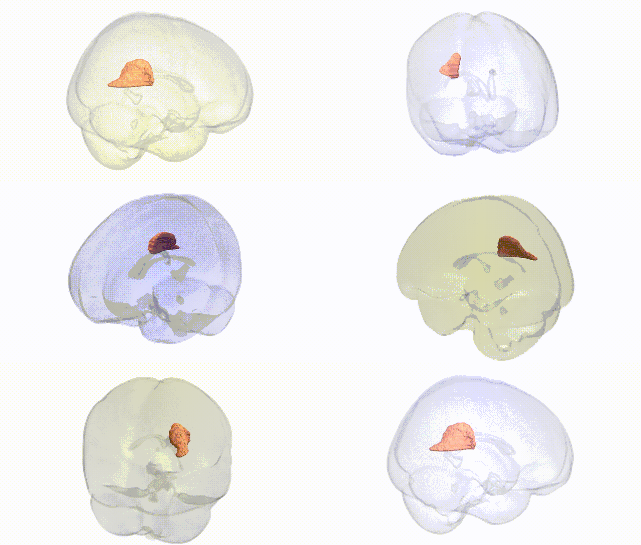
Full Quality Version: Download MP4
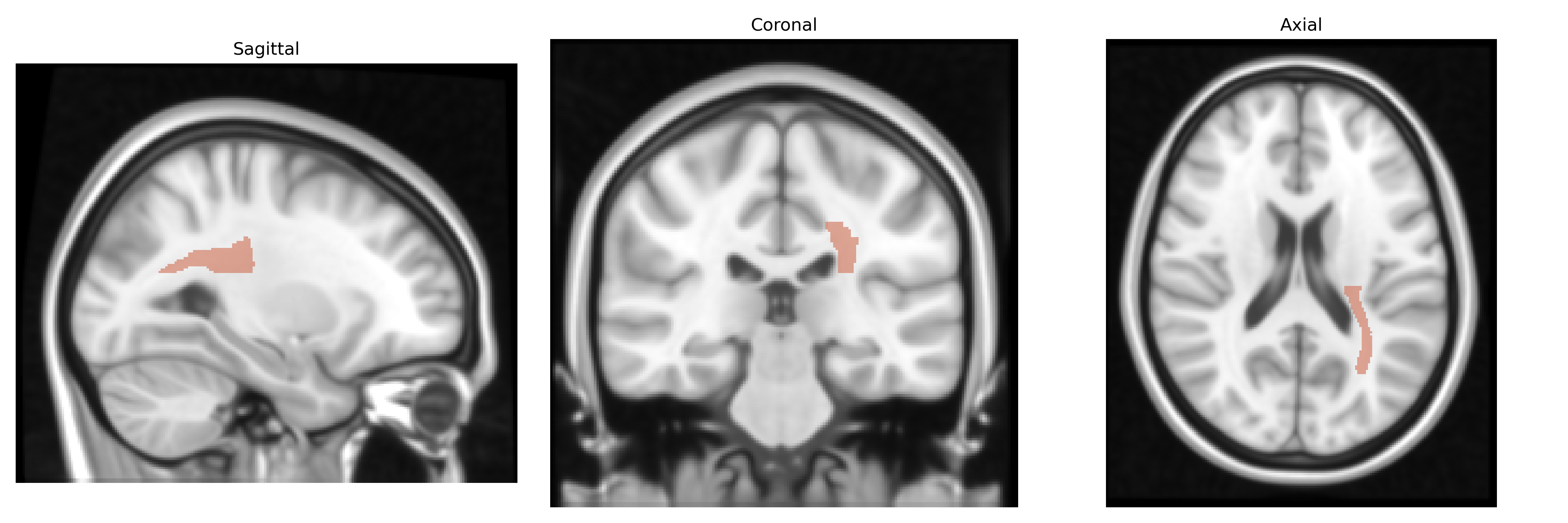
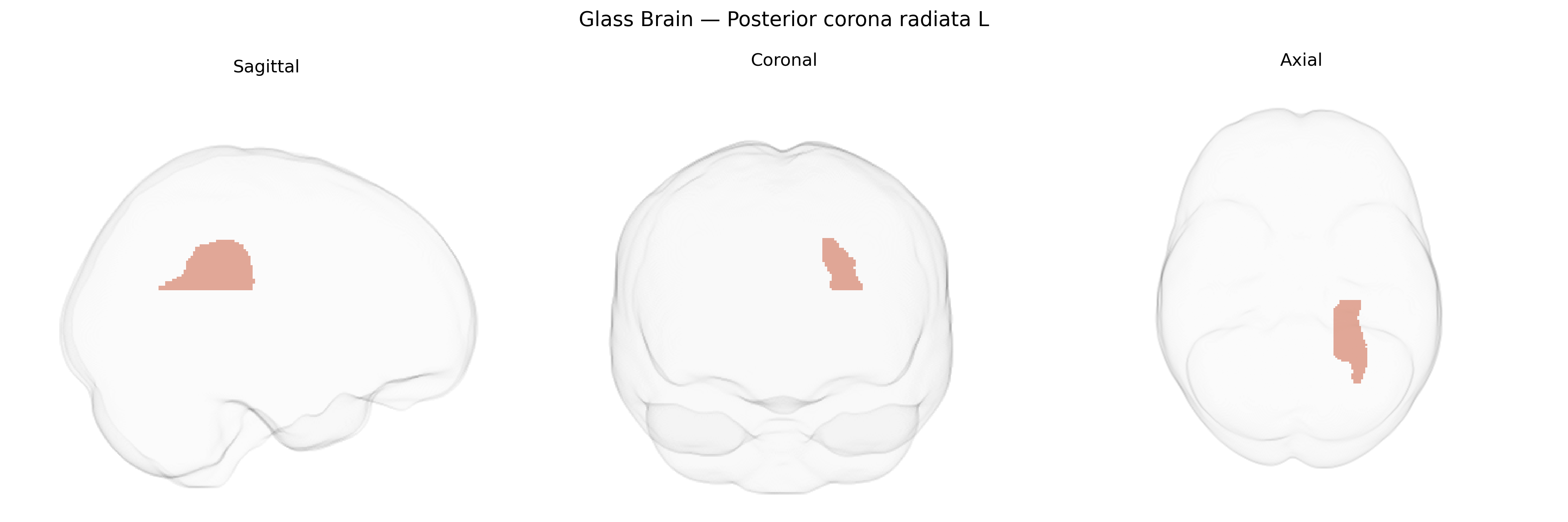
Wali Sidiqyar*, Gaurav Rudravaram*, Elyssa M. McMaster, Trent M. Schwartz, Adam M. Saunders, Kurt G. Schilling, Bennett A. Landman "Introducing SPINS: A Shared Public Visualization Library of Neuroanatomical Structures." Medical Imaging with Deep Learning- short paper
This resource is licensed under CC0 1.0 Universal (Public Domain).