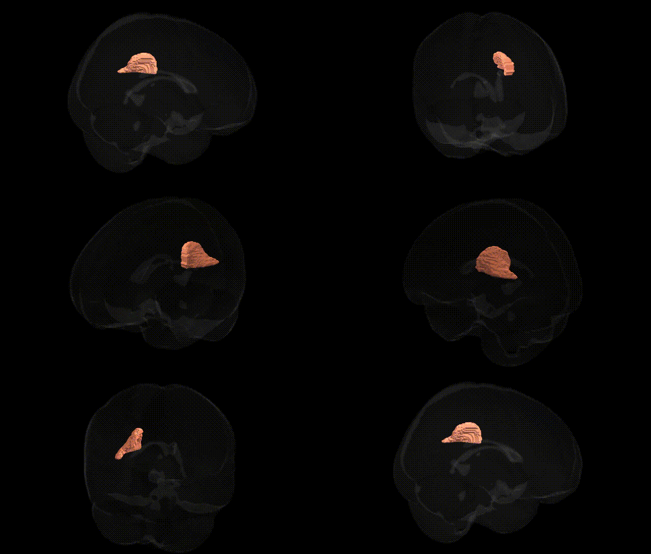
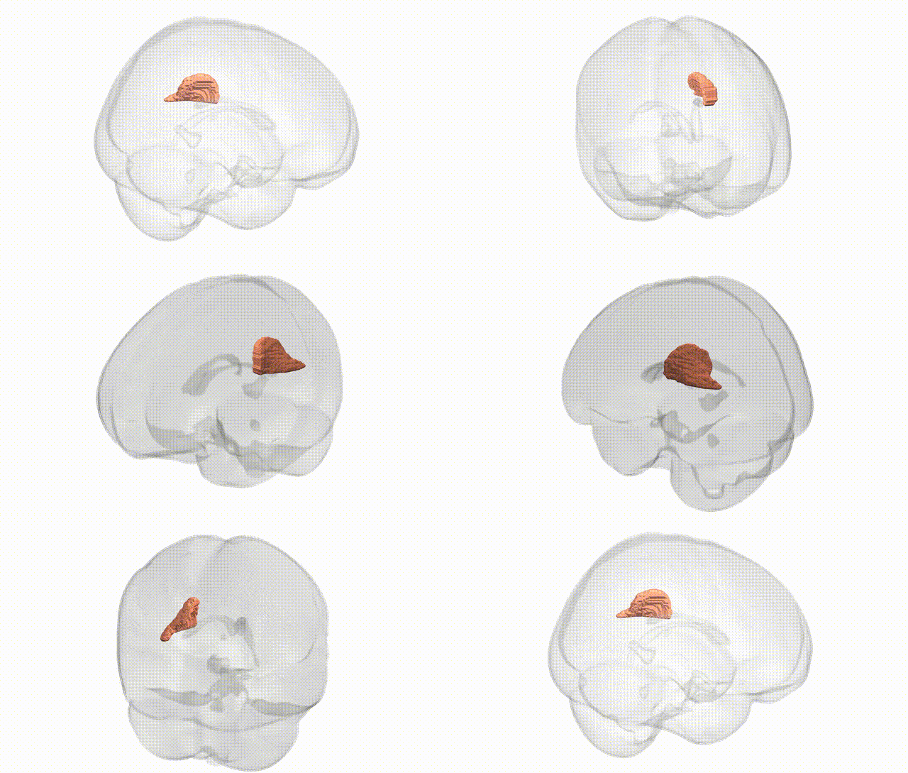
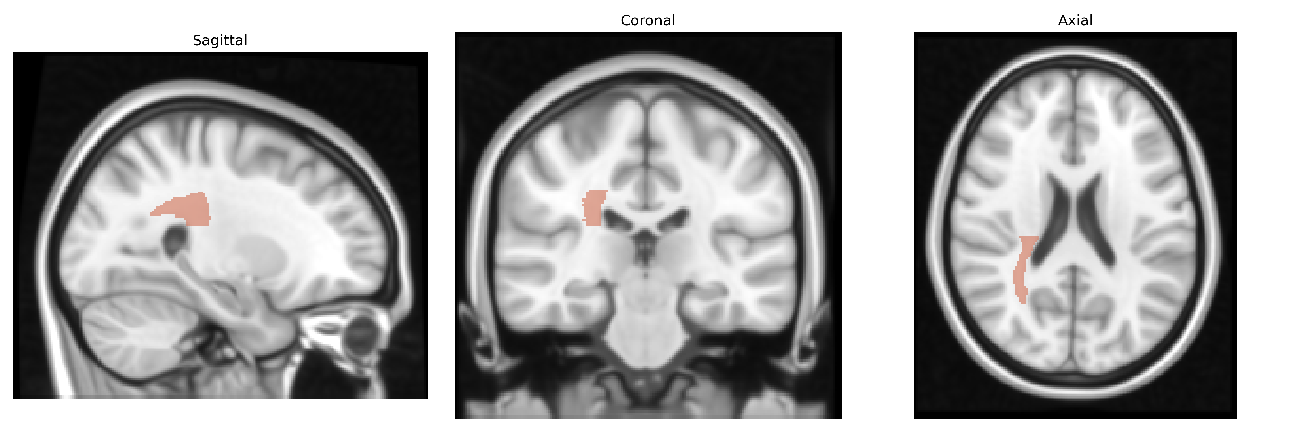
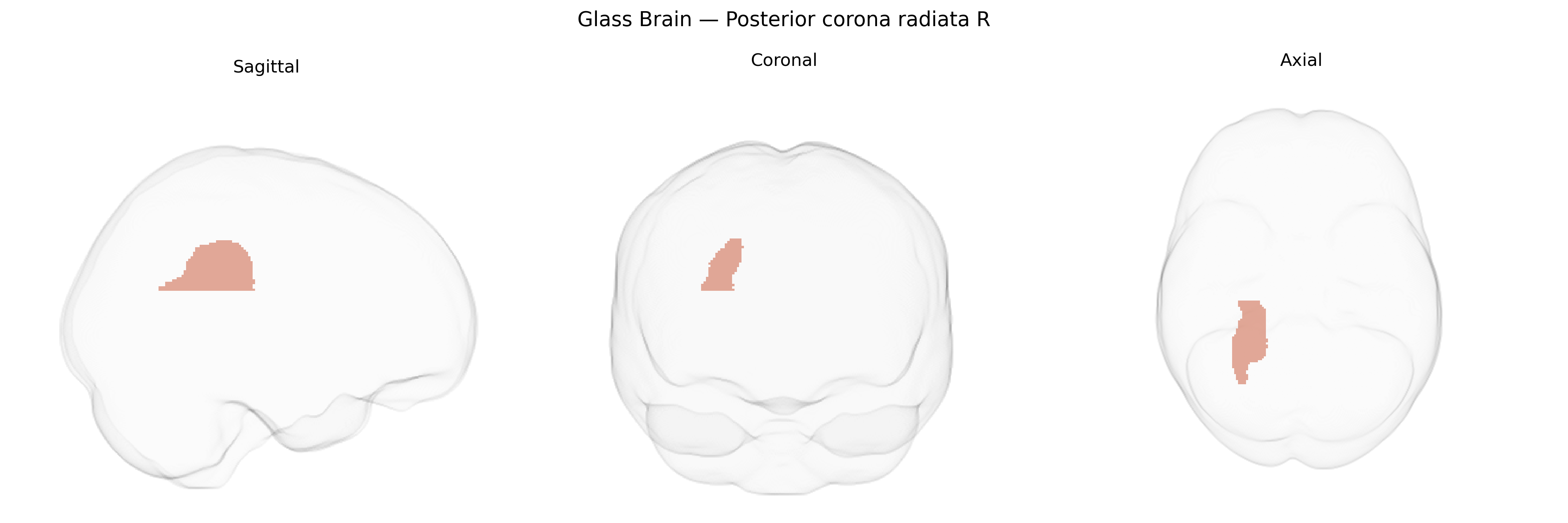
The bilateral Posterior corona radiata R, as defined in the JHU ICBM 1 mm white-matter atlas, corresponds to a segment of the corona radiata located posterior to the central sulcus within the right cerebral hemisphere, comprising predominantly projection fibers that convey sensory and associative information between the cerebral cortex and deeper structures such as the thalamus and brainstem. This region contains fibers from parietal and occipital cortices and contributes to large-scale networks involved in visuospatial processing, somatosensory integration, and higher-order cognitive functions, as well as participating in motor-related pathways through its continuity with the internal capsule and more anterior portions of the corona radiata. Lesions in the posterior corona radiata are frequently implicated in deficits of sensory perception, attention, and visual processing, and the region is a common focus of diffusion tensor imaging studies in conditions such as stroke, traumatic brain injury, and demyelinating disease. There is no direct Wikipedia article for the “Posterior corona radiata R”; a related structure is the Corona radiata.
The bilateral posterior corona radiata (PCR) in the JHU ICBM 1 mm atlas is a major white-matter pathway in the parietal region that has been implicated in multiple GWAS of brain imaging phenotypes, particularly studies using diffusion tensor imaging and structural MRI. Large cohorts such as UK Biobank and ENIGMA have identified SNP-based heritability for fractional anisotropy, mean diffusivity, and regional white-matter volume in the corona radiata, with associated loci frequently mapping to genes involved in axon guidance, myelination, and neurodevelopment (for example, genes near neurodevelopmental loci on chromosomes 3, 6, and 17). Variants in or near genes such as BDNF, NRG1, CNTN4/5, and myelin-related loci (e.g., MAG, MBP regions) have been repeatedly linked to white-matter microstructure measures that include or overlap the PCR, though often in combined tracts rather than PCR-specific analyses. GWAS of psychiatric and neurological disorders—schizophrenia, bipolar disorder, major depression, attention-deficit/hyperactivity disorder, and multiple sclerosis—report case–control differences in corona radiata integrity that co-localize with polygenic risk, with imaging–genetics work showing that higher genetic risk for these disorders is associated with altered white-matter properties in the PCR. Cognitive traits such as general intelligence, processing speed, and working memory have also been linked through imaging–genetic analyses to microstructural variation in the posterior corona radiata, suggesting that genetic influences on this tract contribute to interindividual differences in cognition. Although specific PCR-focused GWAS loci are still relatively sparse, converging evidence from polygenic scores, neurodevelopmental gene sets, and multimodal imaging–genetics studies indicates that the bilateral posterior corona radiata is a genetically influenced hub of white-matter connectivity relevant to psychiatric risk, demyelinating disease, and cognitive performance.
Overview generated by GPT-4o (2026).
Region ID: 27
Hemisphere: bilateral
Atlas: JHU ICBM labels 1mm
Full Quality Version: Download MP4
Full Quality Version: Download MP4
Wali Sidiqyar*, Gaurav Rudravaram*, Elyssa M. McMaster, Trent M. Schwartz, Adam M. Saunders, Kurt G. Schilling, Bennett A. Landman "Introducing SPINS: A Shared Public Visualization Library of Neuroanatomical Structures." Medical Imaging with Deep Learning- short paper
This resource is licensed under CC0 1.0 Universal (Public Domain).