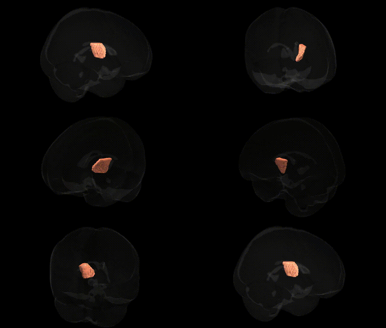
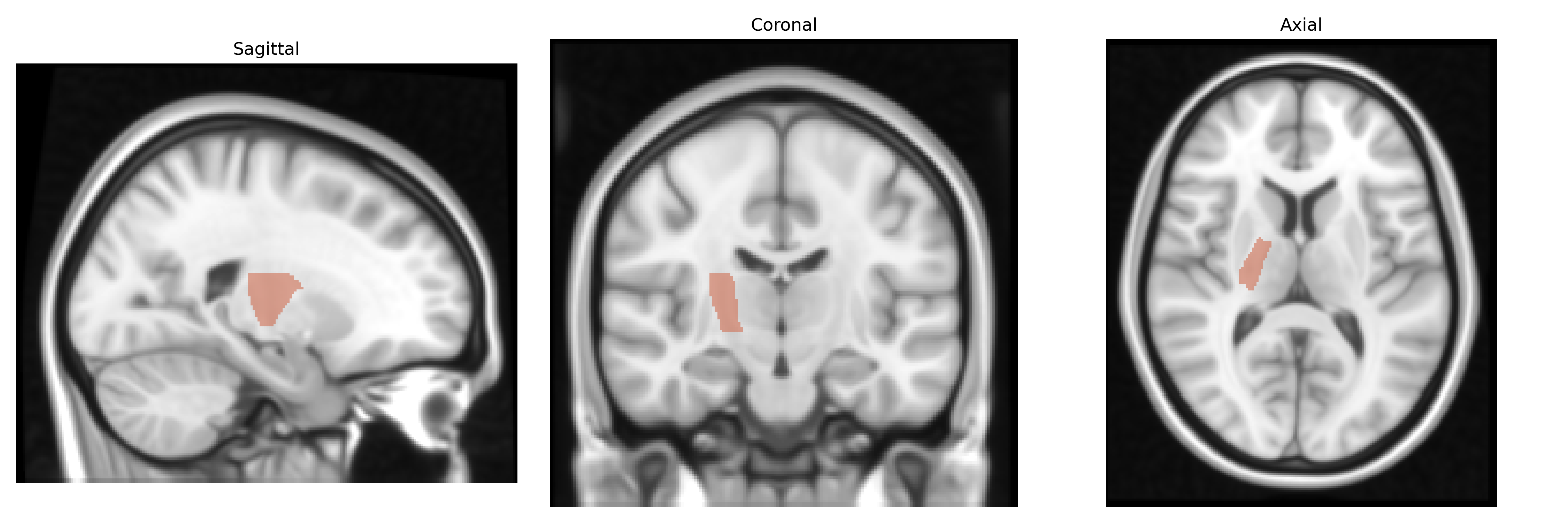
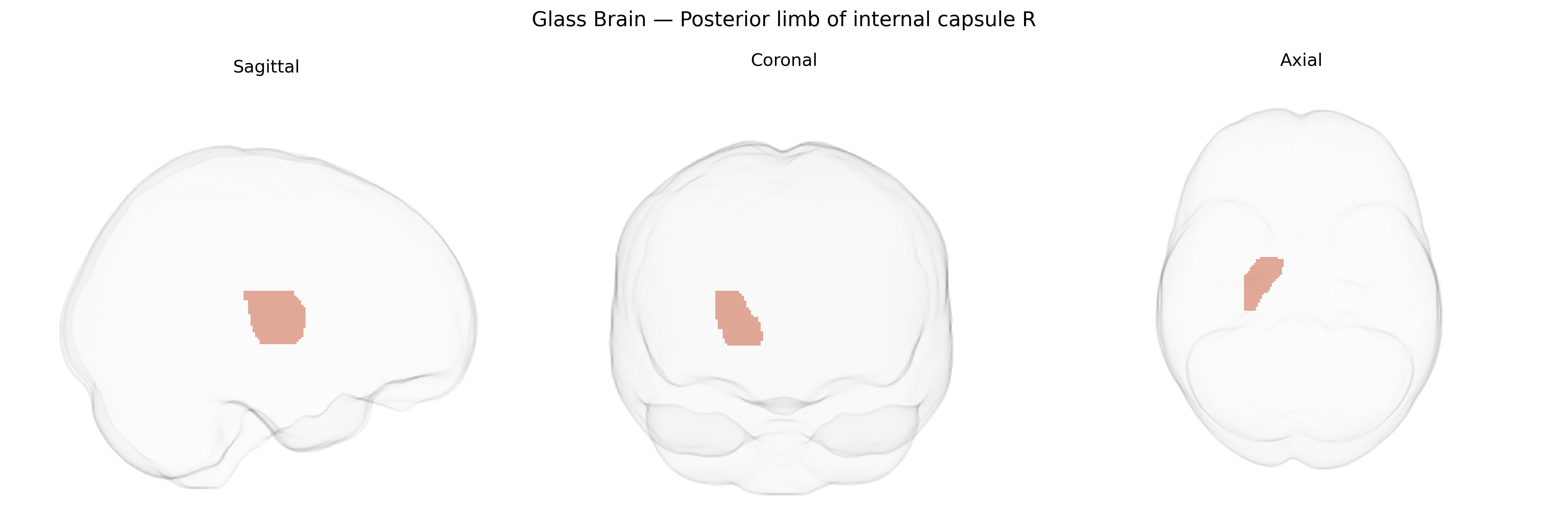
The bilateral posterior limb of the internal capsule (right) is a major white matter conduit in the deep telencephalon that carries descending corticospinal, corticobulbar, and corticopontine fibers, as well as ascending thalamocortical projections, particularly somatosensory pathways from the ventral posterior thalamic nuclei to the primary somatosensory cortex. Positioned between the thalamus medially and the lentiform nucleus laterally, this segment of the internal capsule is somatotopically organized, with fibers representing different body parts arranged in a precise medial–lateral gradient. It plays a critical role in voluntary motor control, somatosensation, and integration of cortical and subcortical communication; lesions here commonly result in contralateral motor weakness, sensory loss, or pure motor strokes due to disruption of compact fiber tracts. There is no direct link for the specific “posterior limb of internal capsule, right” label; see the related structure Internal capsule.
The bilateral posterior limb of the internal capsule (PLIC), as defined in the JHU ICBM 1 mm atlas, has been implicated in multiple neuroimaging genetics studies, largely through diffusion MRI–based GWAS of white matter microstructure. Variants in genes related to axonal guidance, myelination, and oligodendrocyte function—such as those in the neuregulin–ERBB pathway, CNTN4/CNTN6, and myelin-associated genes—have been associated with fractional anisotropy and mean diffusivity in the PLIC, reflecting its dense corticospinal and thalamocortical fiber composition. Large-scale consortia (e.g., ENIGMA) have reported polygenic influences on PLIC integrity, with shared genetic architecture across motor and sensory tracts, and loci near genes involved in neurodevelopment and cytoskeletal organization showing replicated effects. Clinically, genetic variants that increase risk for disorders affecting motor pathways or white matter, including cerebral small vessel disease, multiple sclerosis, and some monogenic leukodystrophies, often show secondary effects on PLIC structure and function, and GWAS signals for ischemic stroke and white matter hyperintensities include regions that intersect the PLIC. Additionally, polygenic risk for conditions such as schizophrenia, bipolar disorder, and major depression has been linked to altered PLIC microstructure in imaging–genetics studies, suggesting pleiotropic genetic influences on this tract, although specific locus-to-region mappings remain largely probabilistic rather than region-specific.
Overview generated by GPT-4o (2026).
Region ID: 19
Hemisphere: bilateral
Atlas: JHU ICBM labels 1mm
Full Quality Version: Download MP4
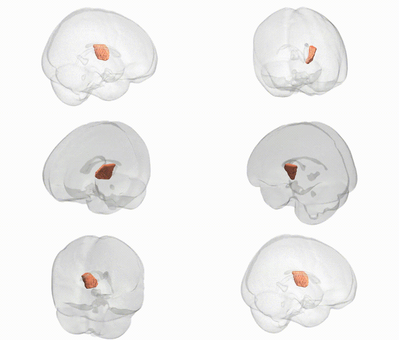
Full Quality Version: Download MP4
Wali Sidiqyar*, Gaurav Rudravaram*, Elyssa M. McMaster, Trent M. Schwartz, Adam M. Saunders, Kurt G. Schilling, Bennett A. Landman "Introducing SPINS: A Shared Public Visualization Library of Neuroanatomical Structures." Medical Imaging with Deep Learning- short paper
This resource is licensed under CC0 1.0 Universal (Public Domain).