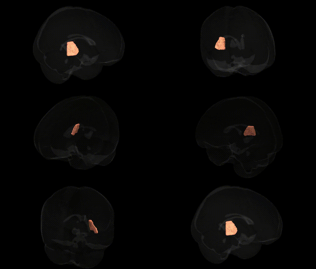
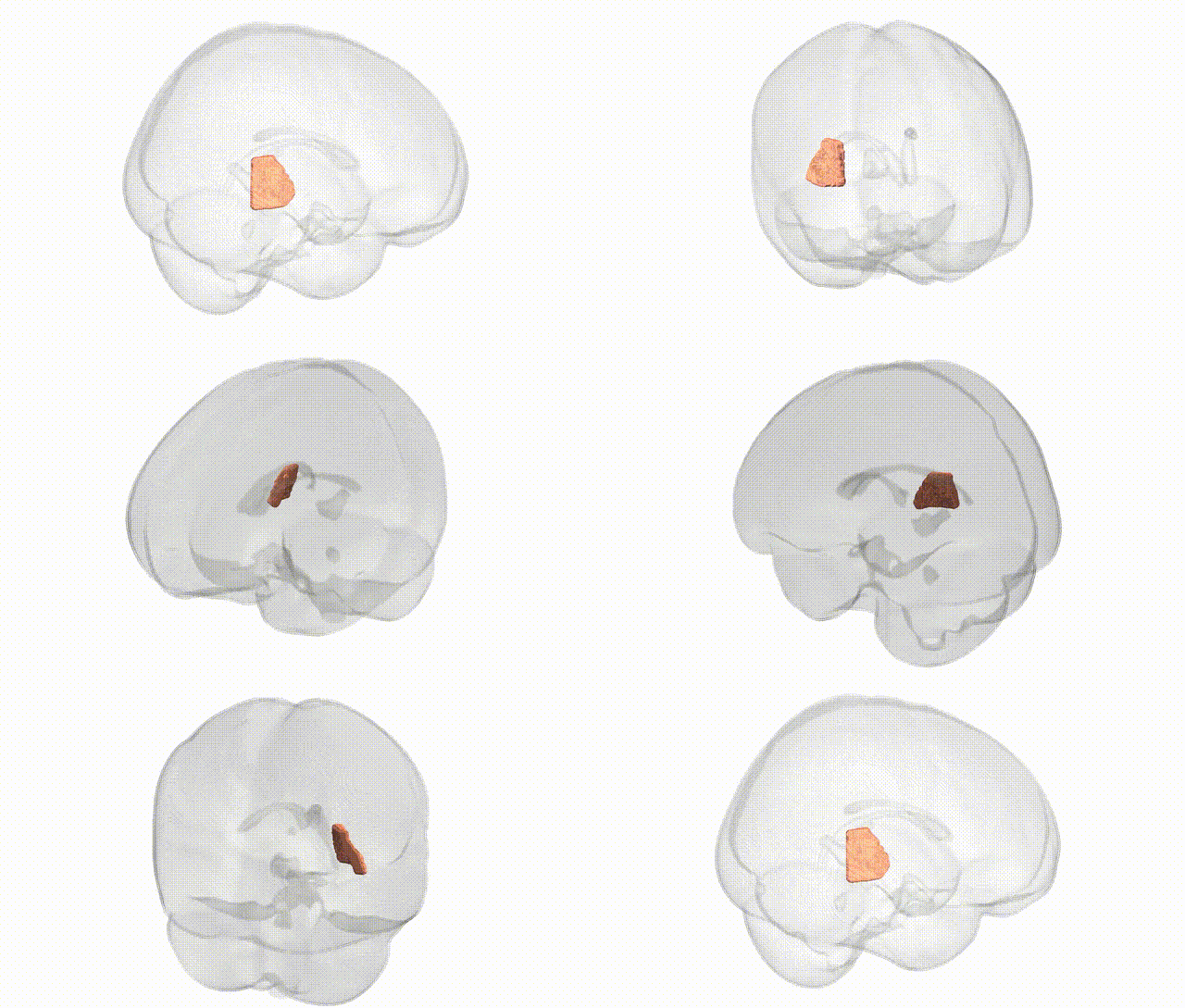
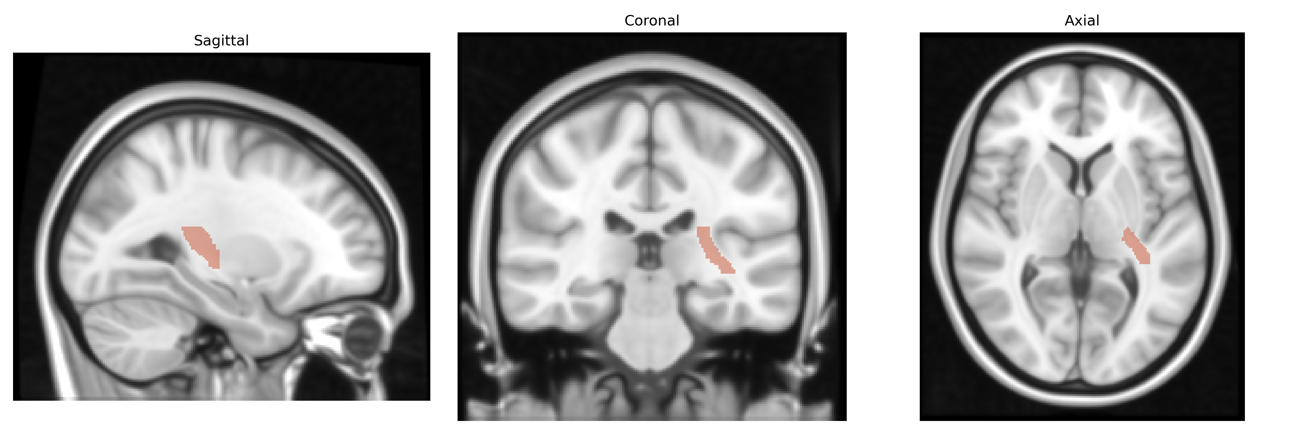
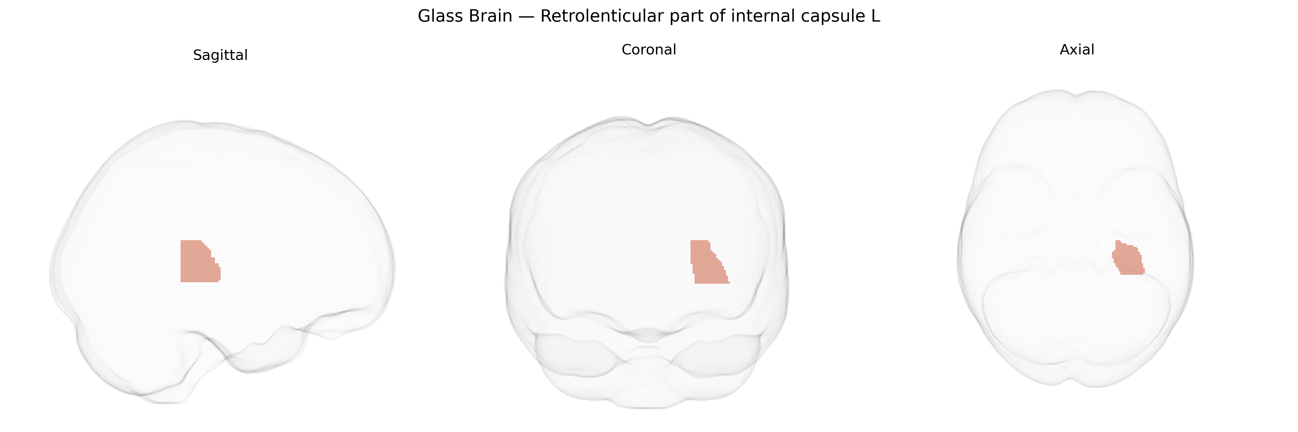
The bilateral retrolenticular part of the internal capsule (left) is a posterior segment of the internal capsule situated behind the lentiform nucleus, containing major projection fibers that connect the thalamus and cerebral cortex. It primarily carries the optic radiation (geniculocalcarine tract) from the lateral geniculate nucleus to the primary visual cortex, as well as auditory radiations and some parietal association fibers, thereby playing a critical role in visual and auditory processing and higher-order sensory integration. Damage to this region can lead to visual field deficits (such as homonymous hemianopia) and disruptions in sensory perception and cortical communication. There is no direct Wikipedia article for this specific subregion; a related structure is the Internal capsule.
Genetic associations specifically targeting the bilateral retrolenticular part of the internal capsule (RL-IC) in the JHU ICBM 1 mm atlas are limited, but several lines of evidence implicate this region within broader white-matter and subcortical connectivity phenotypes. Large imaging-genetics GWAS using diffusion MRI (e.g., UK Biobank–based studies) have identified heritable variation in fractional anisotropy, mean diffusivity, and tract integrity in posterior limb and retrolenticular regions of the internal capsule linked to loci such as those near CNTN4, COL4A2, and genes involved in axon guidance, myelination (e.g., MAG, MBP), and vascular integrity. White-matter microstructure in internal capsule tracts, including retrolenticular fibers projecting to parietal and occipital cortices, has been associated via polygenic risk scores and GWAS hits with neuropsychiatric and neurodevelopmental disorders (schizophrenia, bipolar disorder, major depression, ADHD, autism), cognitive traits (general intelligence, processing speed), and cerebrovascular phenotypes (small-vessel disease, ischemic stroke), with risk loci such as those in CACNA1C, ZNF804A, and other synaptic/neuronal genes influencing tract-specific metrics. Additionally, rare variants and Mendelian disorders affecting myelin and axonal maintenance (e.g., leukodystrophies with mutations in PLP1, EIF2B genes) often show prominent involvement of internal capsule white matter, including retrolenticular portions, underscoring a genetically mediated vulnerability of this region. However, most genetic findings to date treat the retrolenticular internal capsule as part of composite internal capsule or global white-matter measures rather than as an isolated atlas-defined structure, so region-specific GWAS signals are inferred from tract-wide or voxelwise analyses rather than focused locus-phenotype studies.
Overview generated by GPT-4o (2026).
Region ID: 22
Hemisphere: bilateral
Atlas: JHU ICBM labels 1mm
Full Quality Version: Download MP4
Full Quality Version: Download MP4
Wali Sidiqyar*, Gaurav Rudravaram*, Elyssa M. McMaster, Trent M. Schwartz, Adam M. Saunders, Kurt G. Schilling, Bennett A. Landman "Introducing SPINS: A Shared Public Visualization Library of Neuroanatomical Structures." Medical Imaging with Deep Learning- short paper
This resource is licensed under CC0 1.0 Universal (Public Domain).