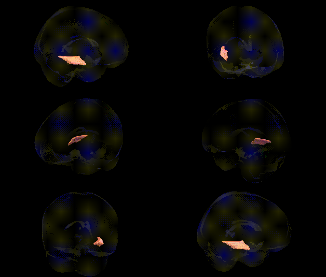
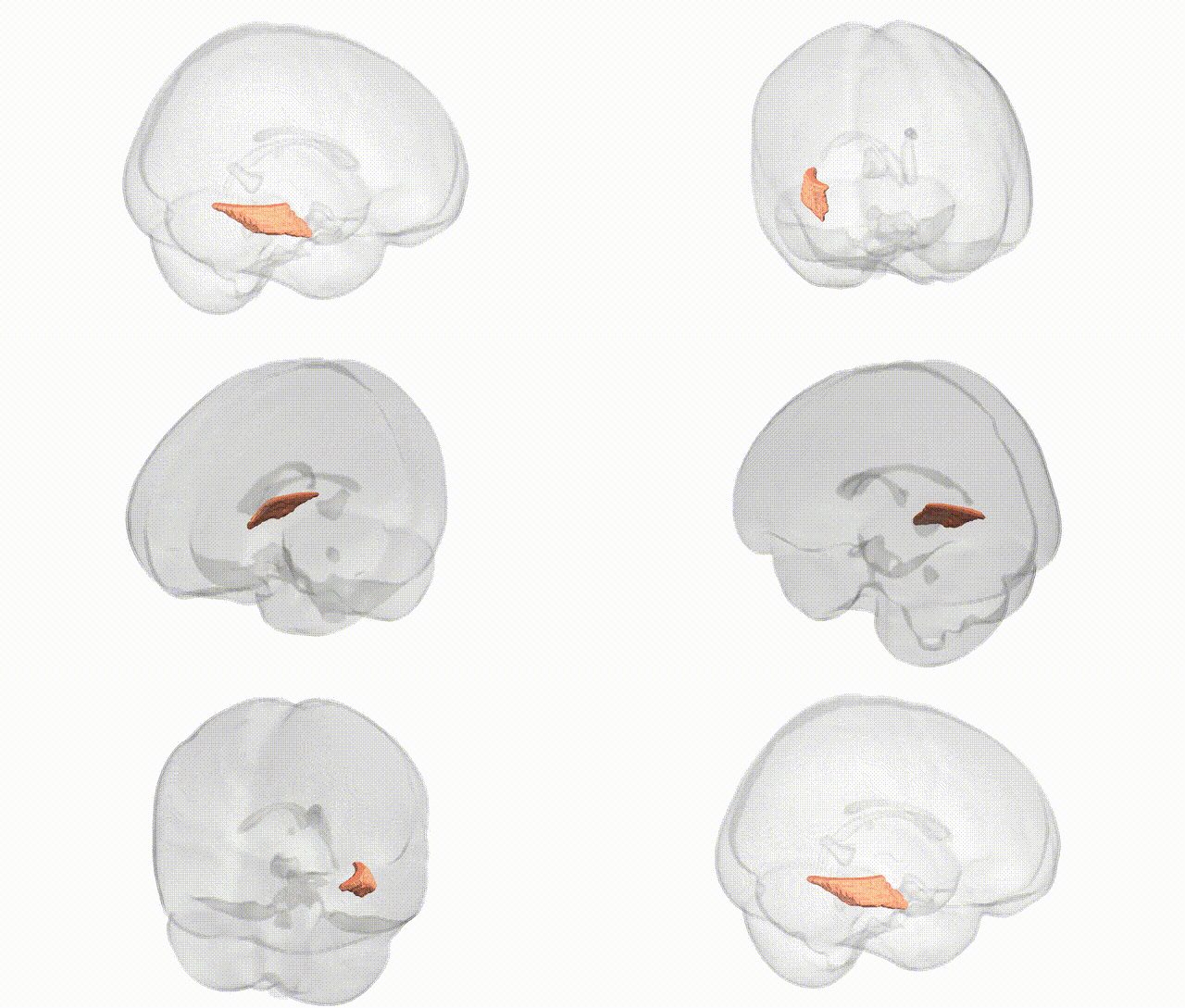
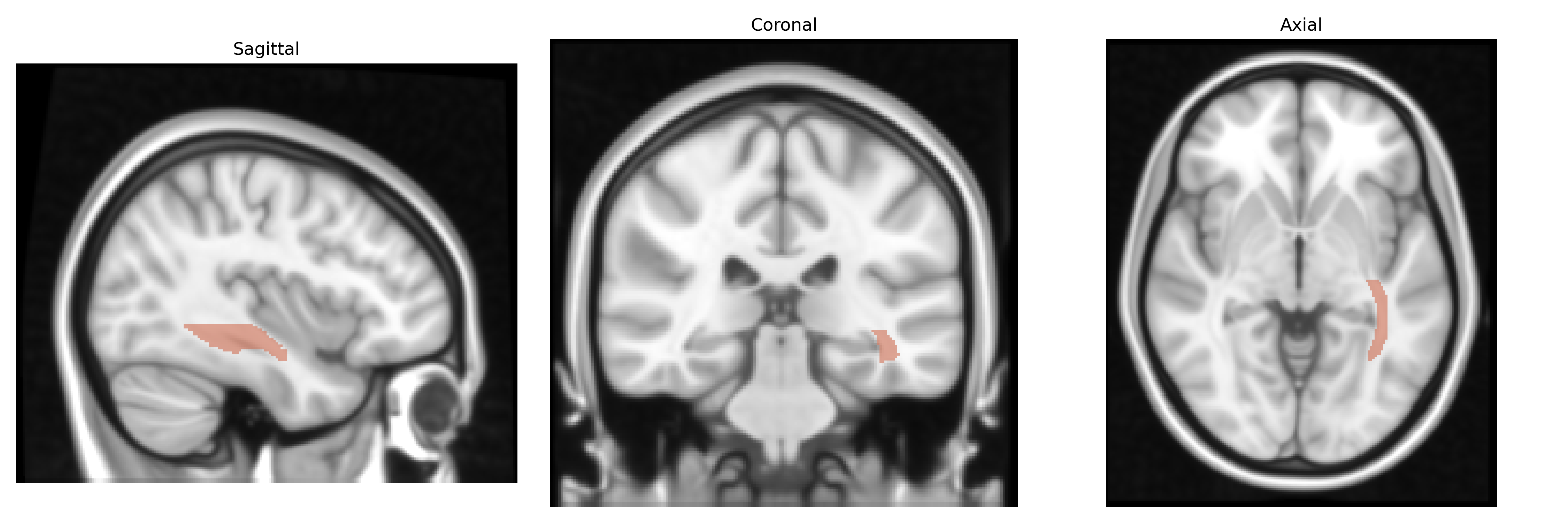
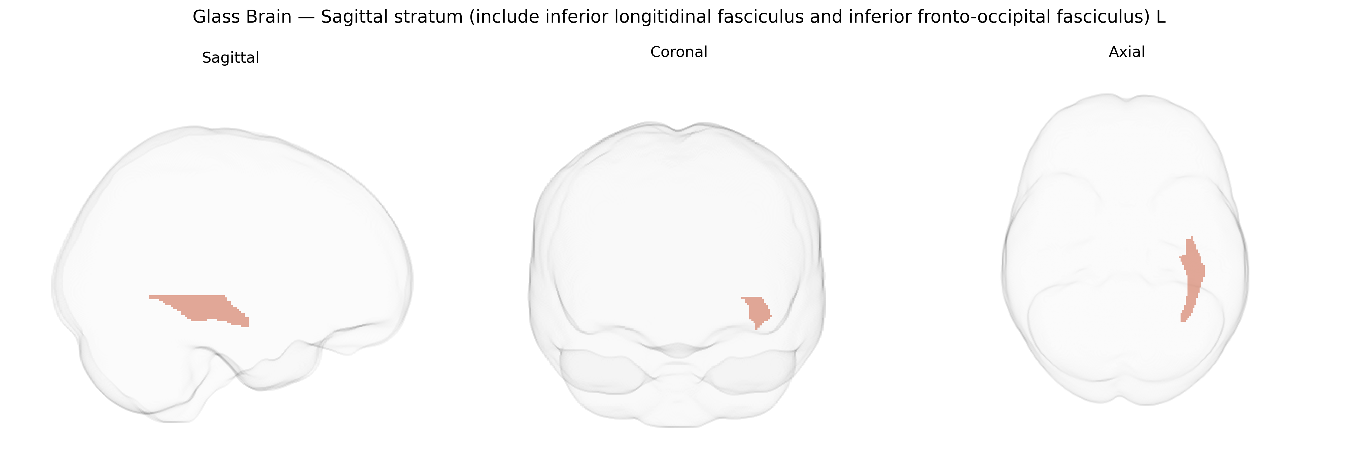
The bilateral sagittal stratum (including the inferior longitudinal fasciculus and inferior fronto-occipital fasciculus) L, as defined in the JHU ICBM labels 1 mm atlas, is a major composite white matter region situated in the deep posterior temporal and occipital lobes, forming part of the ventral associative pathways that connect occipital, temporal, and frontal cortices. The inferior longitudinal fasciculus primarily links occipital visual regions with anterior and medial temporal structures, supporting visual object recognition and aspects of semantic and memory processing, while the inferior fronto-occipital fasciculus courses from occipital and posterior temporal areas to the frontal lobe, contributing to higher-order visual integration, language, and executive functions. Together within the sagittal stratum, these tracts carry long-range cortico-cortical fibers that integrate multimodal sensory information with cognitive and linguistic processes, and are commonly studied in diffusion MRI for their role in large-scale brain networks and in disorders affecting white matter integrity. There is no direct Wikipedia article for the sagittal stratum as defined in this atlas; a closely related structure is the Inferior longitudinal fasciculus.
The bilateral sagittal stratum (including the inferior longitudinal fasciculus and inferior fronto-occipital fasciculus) as defined in the JHU ICBM 1 mm atlas has repeatedly emerged in imaging‑genetics and GWAS of white‑matter microstructure, where diffusion parameters such as fractional anisotropy and mean diffusivity in these tracts show substantial SNP‑based heritability and polygenic influences. Large consortia (e.g., ENIGMA‑DTI, UK Biobank imaging GWAS) have identified common variants in genes involved in axonal guidance, myelination, and oligodendrocyte function (such as NTRK1/2, MAG, and other myelin‑related loci) that are associated with diffusion metrics in the inferior longitudinal and inferior fronto‑occipital fasciculi, though specific locus–tract assignments are often reported at the level of broader temporal–occipital or association white matter rather than this atlas label alone. Polygenic risk scores for schizophrenia, bipolar disorder, major depressive disorder, and autism spectrum disorder have been linked to reduced integrity of these tracts, and case–control imaging‑genetic studies indicate that risk variants in genes such as ZNF804A, CACNA1C, and neuregulin/ERBB signaling components may modulate connectivity between occipital, temporal, and frontal regions via effects on sagittal stratum pathways. Rare copy‑number variants and Mendelian mutations impacting myelin (for example, in leukodystrophies and some forms of hereditary spastic paraplegia) often produce marked abnormalities in this region on MRI, reinforcing its vulnerability to genetic disruption of white‑matter development. Collectively, evidence supports a polygenic architecture in which variants influencing neurodevelopment, synaptic plasticity, and myelin biology contribute to interindividual differences and disease‑related alterations in the bilateral sagittal stratum and its constituent tracts.
Overview generated by GPT-4o (2026).
Region ID: 32
Hemisphere: bilateral
Atlas: JHU ICBM labels 1mm
Full Quality Version: Download MP4
Full Quality Version: Download MP4
Wali Sidiqyar*, Gaurav Rudravaram*, Elyssa M. McMaster, Trent M. Schwartz, Adam M. Saunders, Kurt G. Schilling, Bennett A. Landman "Introducing SPINS: A Shared Public Visualization Library of Neuroanatomical Structures." Medical Imaging with Deep Learning- short paper
This resource is licensed under CC0 1.0 Universal (Public Domain).