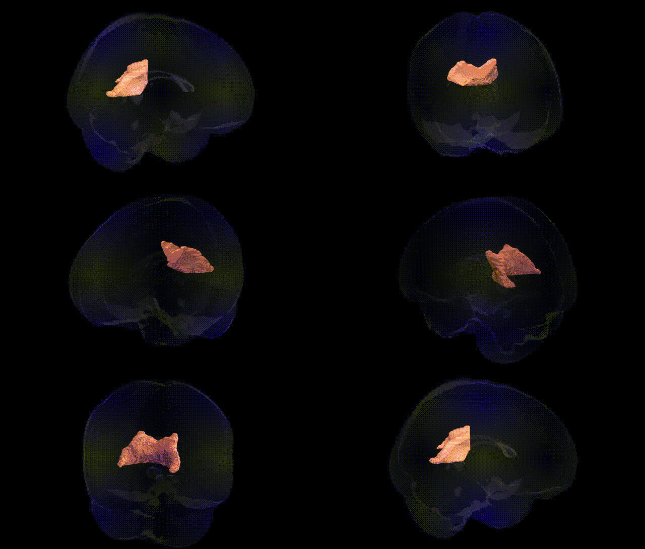
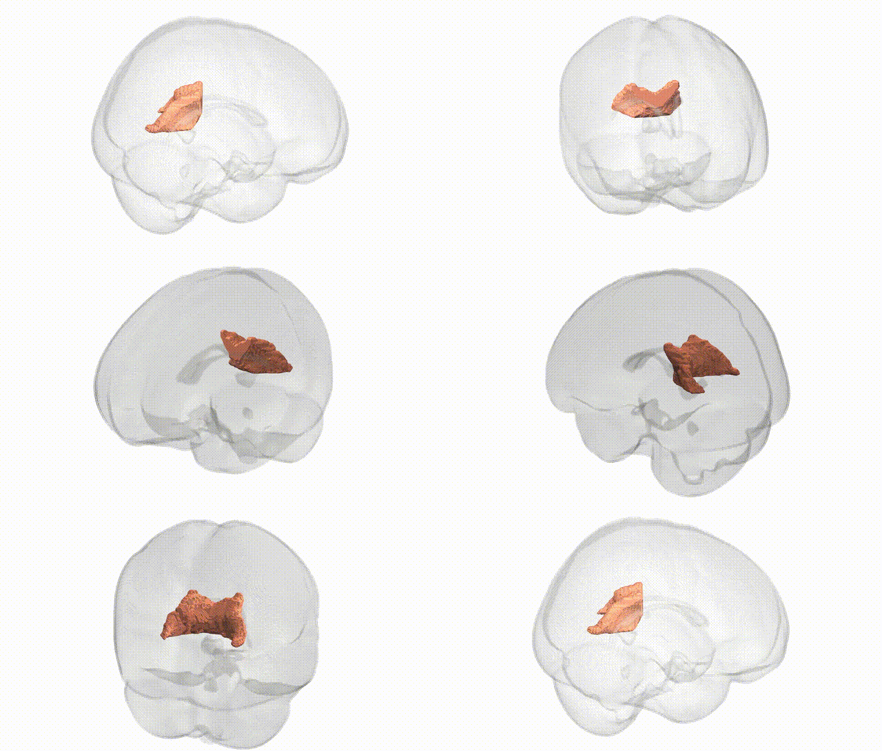
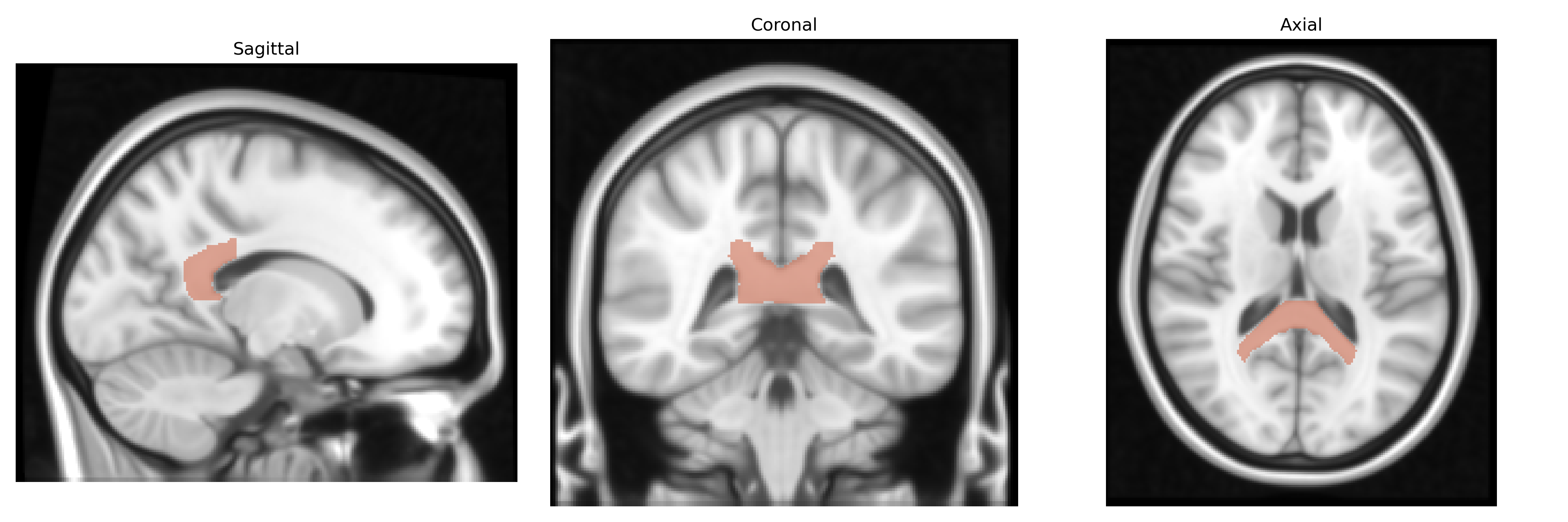
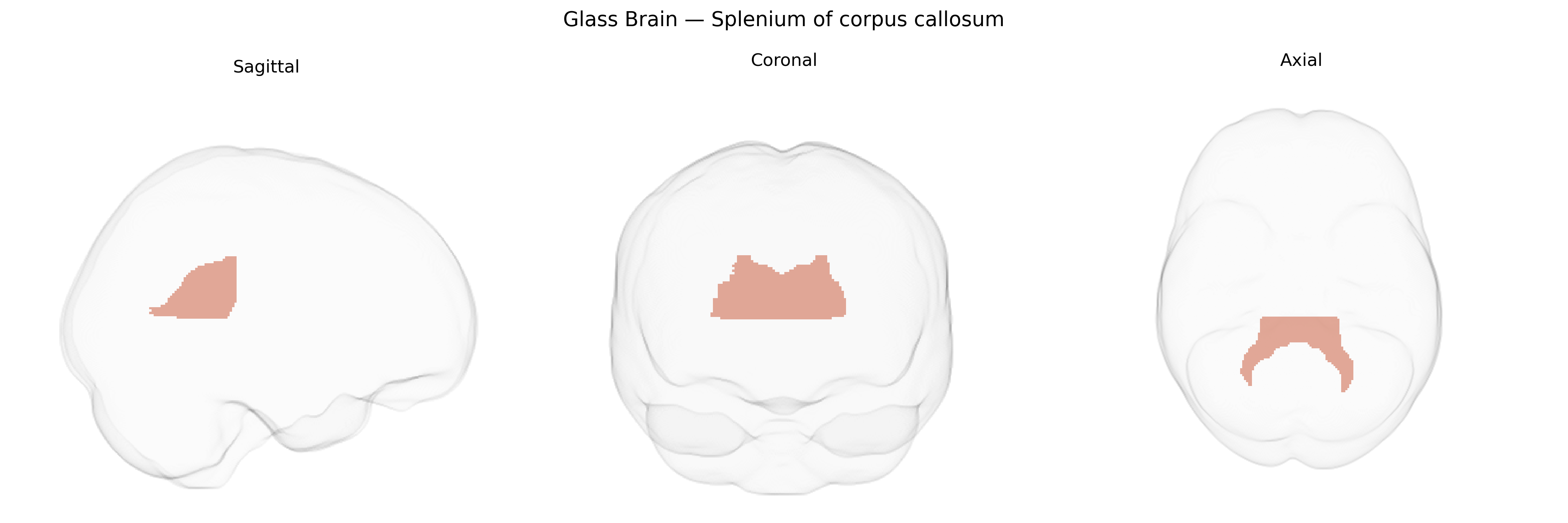
The bilateral splenium of the corpus callosum is the posterior portion of the corpus callosum, comprising densely packed commissural fibers that interconnect homologous regions of the occipital, posterior parietal, and temporo-occipital cortices across the two cerebral hemispheres. In the JHU ICBM labels 1 mm Atlas, this region is delineated as a major white matter structure critical for interhemispheric transfer of visual and visuospatial information, as well as higher-order multimodal integration. The splenium’s robust myelination supports rapid conduction of signals essential for coordinated bilateral visual processing, reading, and aspects of language and memory that rely on posterior cortical networks. There is no direct Wikipedia article for the “splenium of corpus callosum” alone; it is described within the broader structure of the Corpus callosum.
The bilateral splenium of the corpus callosum, as defined in the JHU ICBM labels 1 mm atlas, has been implicated in multiple genetic and GWAS findings, largely through studies of white matter microstructure (e.g., fractional anisotropy and mean diffusivity) rather than volume alone. Variants in genes involved in axon guidance, myelination, and oligodendrocyte function—such as CNTNAP2, NRG1, MAG, and myelin-related loci identified in large diffusion MRI GWAS (including polygenic influences across many loci like those near DPYSL2 and VCAN)—have been associated with interindividual differences in callosal integrity, with the splenium often highlighted due to its dense posterior fiber tracts. GWAS and candidate gene studies in schizophrenia, bipolar disorder, and major depressive disorder have linked risk alleles (e.g., in ZNF804A, DISC1, and CACNA1C) to altered splenial diffusion metrics or callosal morphology, suggesting genetically mediated vulnerability of this tract in psychosis and mood disorders. Neurodevelopmental conditions such as autism spectrum disorder and ADHD show splenial abnormalities tied to risk variants in synaptic and neurodevelopmental genes (e.g., CNTNAP2, AUTS2) in imaging-genetics work, while APOE ε4 and other Alzheimer’s disease risk loci have been associated with accelerated microstructural decline in the splenium, reflecting genetically influenced vulnerability to neurodegeneration. Polygenic scores for cognitive performance and educational attainment also correlate with splenial integrity, consistent with a highly distributed genetic architecture in which many small-effect variants contribute to posterior callosal connectivity that supports visual, language, and higher-order cognitive networks.
Overview generated by GPT-4o (2026).
Region ID: 5
Hemisphere: bilateral
Atlas: JHU ICBM labels 1mm
Full Quality Version: Download MP4
Full Quality Version: Download MP4
Wali Sidiqyar*, Gaurav Rudravaram*, Elyssa M. McMaster, Trent M. Schwartz, Adam M. Saunders, Kurt G. Schilling, Bennett A. Landman "Introducing SPINS: A Shared Public Visualization Library of Neuroanatomical Structures." Medical Imaging with Deep Learning- short paper
This resource is licensed under CC0 1.0 Universal (Public Domain).