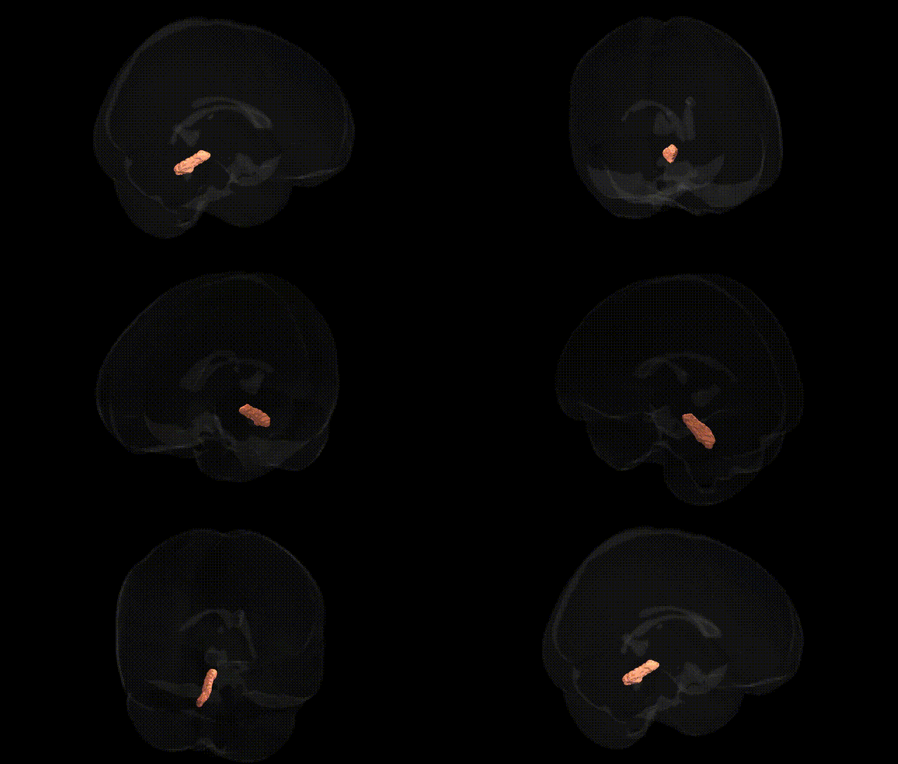
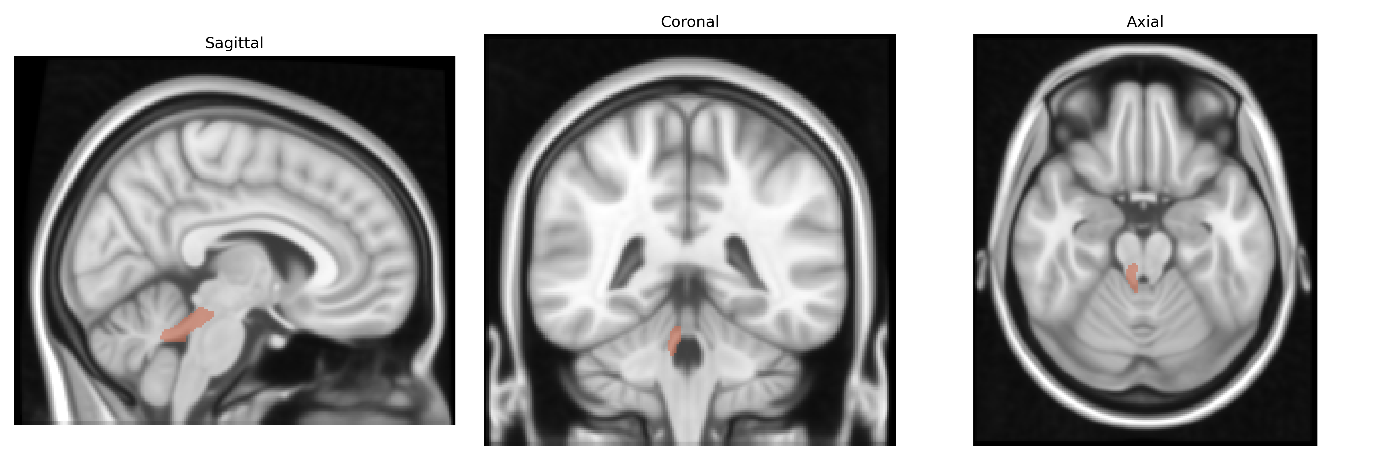
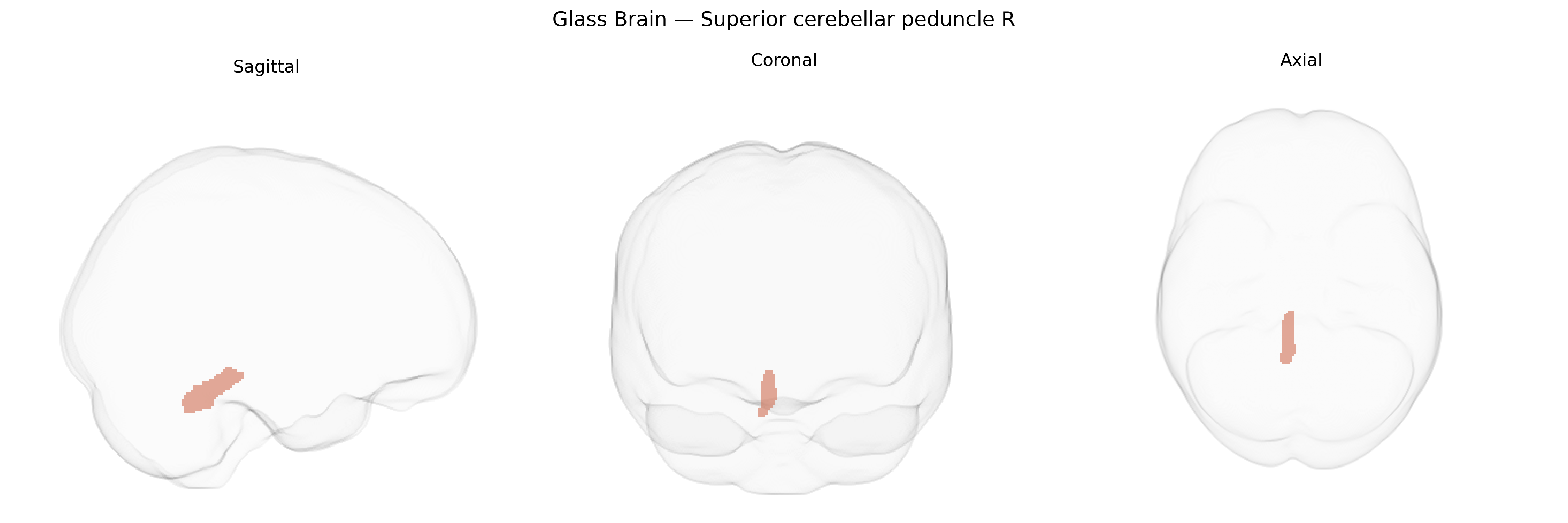
The bilateral Superior cerebellar peduncle (right) is a major efferent white matter tract connecting the cerebellum to the midbrain, and through it to thalamic and cerebral cortical motor areas. Composed largely of fibers arising from the deep cerebellar nuclei (especially the dentate nucleus), it carries processed cerebellar output involved in coordination, timing, and precision of voluntary movement, as well as aspects of motor learning and cognitive modulation of motor behavior. The superior cerebellar peduncle decussates in the caudal midbrain, so right-sided fibers primarily influence contralateral cerebral structures. Lesions affecting this tract can lead to ataxia, dysmetria, and other cerebellar outflow syndromes. There is no direct Wikipedia article for the superior cerebellar peduncle; see the related structure Cerebellar peduncle.
The bilateral superior cerebellar peduncles, as defined in the JHU ICBM 1 mm atlas (right superior cerebellar peduncle R and its left counterpart), have been implicated in genetic studies primarily through imaging genetics and GWAS of brain microstructure rather than direct disease-locus mapping. Large-scale diffusion MRI GWAS (e.g., UK Biobank–based studies) have identified common variants in genes involved in axon guidance, myelination, and neurodevelopment (such as CNTN4, ROBO, and oligodendrocyte-related loci) that influence fractional anisotropy and other diffusion metrics in cerebellar peduncular tracts, including the superior cerebellar peduncle. Polygenic risk for neurodevelopmental and psychiatric conditions—particularly schizophrenia, bipolar disorder, autism spectrum disorder, and ADHD—has been associated with microstructural alterations in cerebellar white matter, and several case–control and endophenotype studies show that risk alleles in genes affecting synaptic function (e.g., CACNA1C, GRM3) and neuronal migration correspond to connectivity changes along cerebello-thalamo-cortical circuits traversing the superior cerebellar peduncle. Additionally, rare variants and copy number changes in genes involved in cerebellar development (e.g., FOXC1, VLDLR, and certain tubulin genes) are linked to congenital cerebellar malformations and ataxia syndromes that typically involve peduncular pathways; while not always mapped voxel-wise to JHU labels, structural and diffusion analyses frequently show superior cerebellar peduncle abnormalities in these conditions. Finally, GWAS of motor coordination, balance, and cognitive traits have reported associations with loci regulating brainstem and cerebellar connectivity, suggesting that genetically driven variation in the superior cerebellar peduncle contributes to individual differences in motor, cognitive, and affective phenotypes, although these links remain largely indirect and mediated via broader cerebello-thalamic networks rather than region-specific risk loci.
Overview generated by GPT-4o (2026).
Region ID: 13
Hemisphere: bilateral
Atlas: JHU ICBM labels 1mm
Full Quality Version: Download MP4
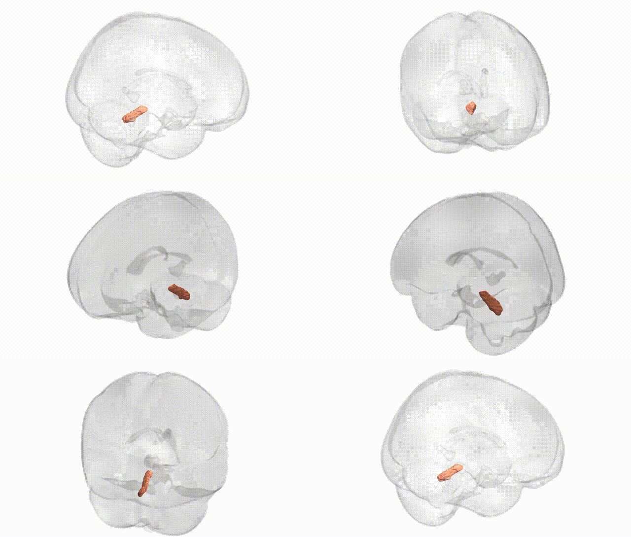
Full Quality Version: Download MP4
Wali Sidiqyar*, Gaurav Rudravaram*, Elyssa M. McMaster, Trent M. Schwartz, Adam M. Saunders, Kurt G. Schilling, Bennett A. Landman "Introducing SPINS: A Shared Public Visualization Library of Neuroanatomical Structures." Medical Imaging with Deep Learning- short paper
This resource is licensed under CC0 1.0 Universal (Public Domain).