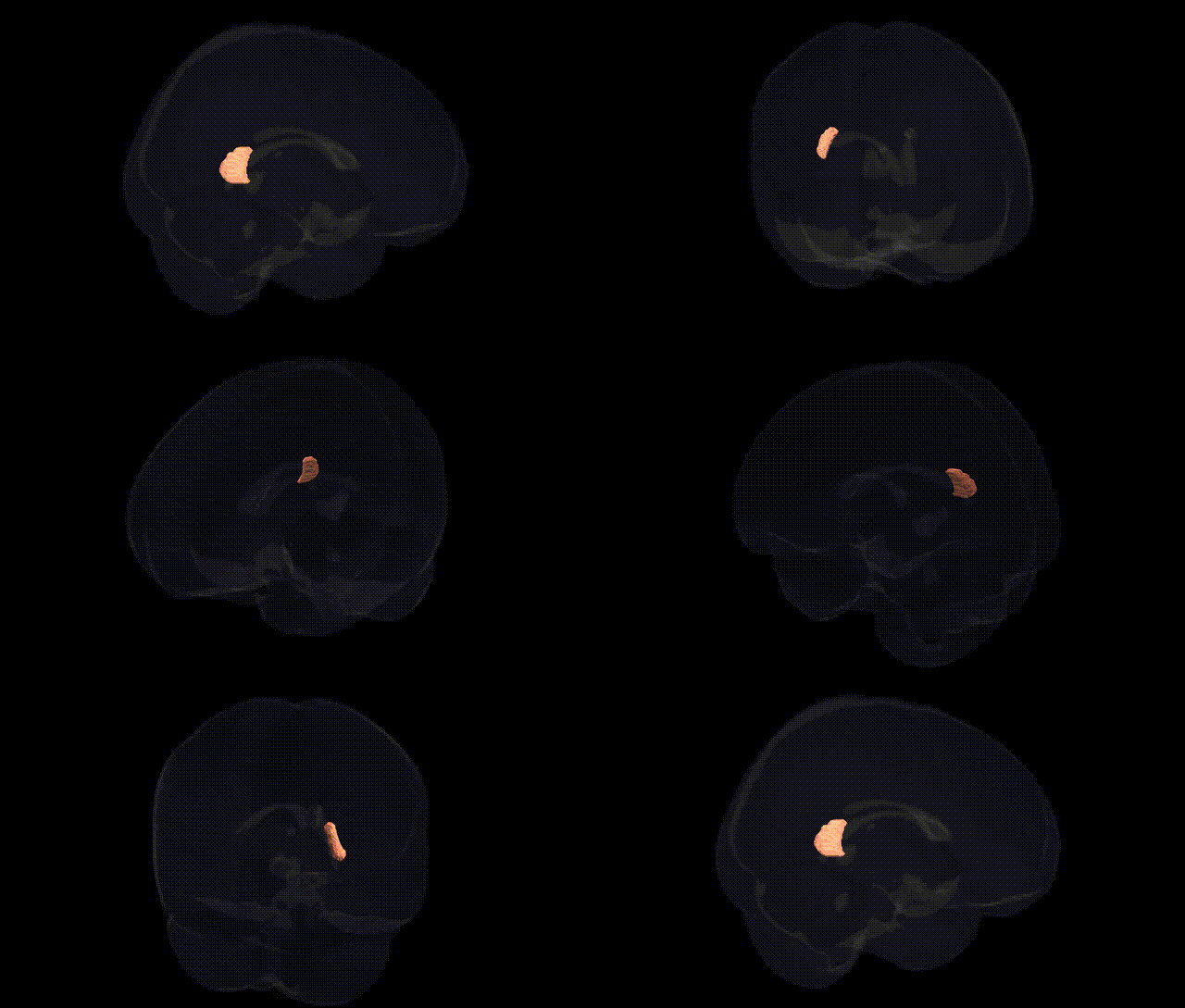
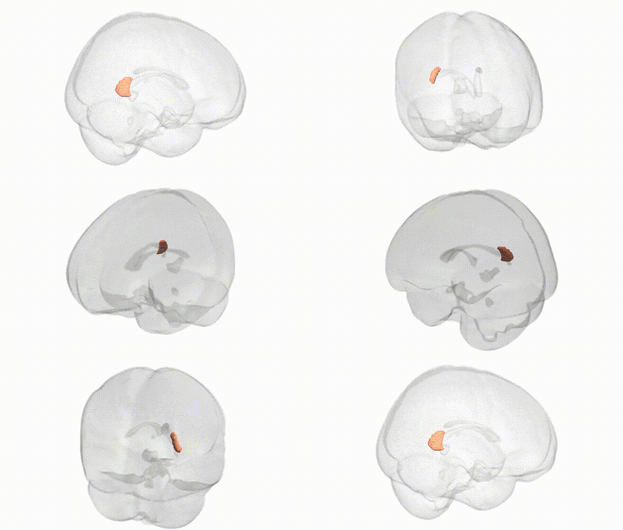
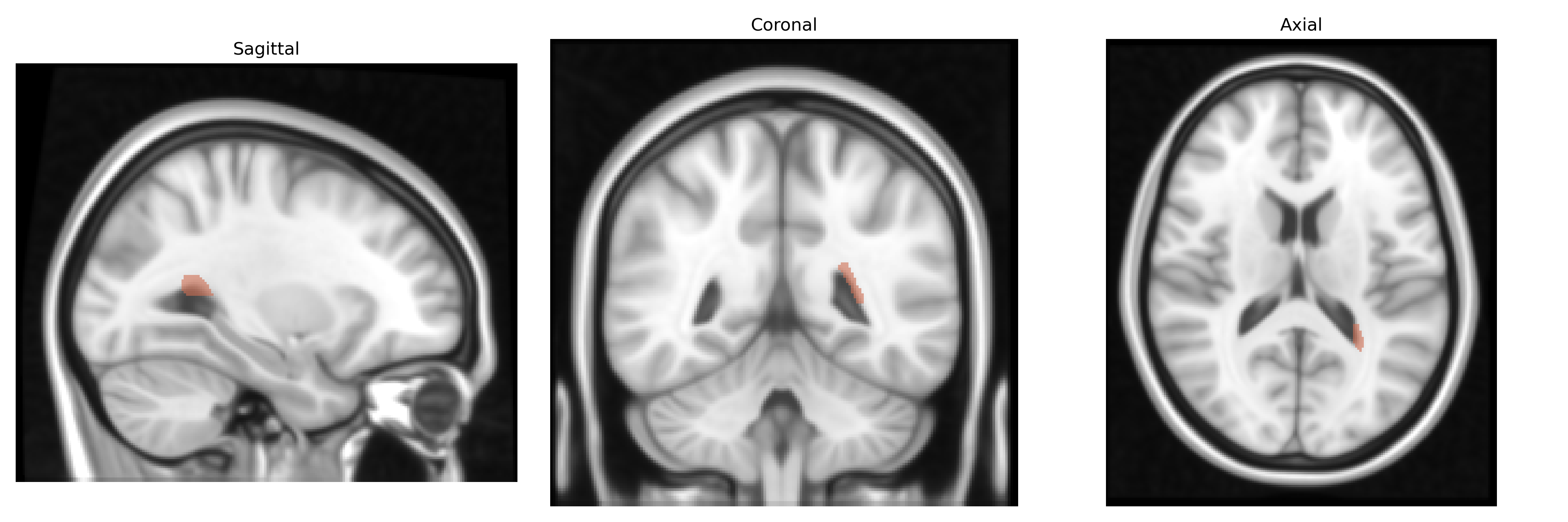
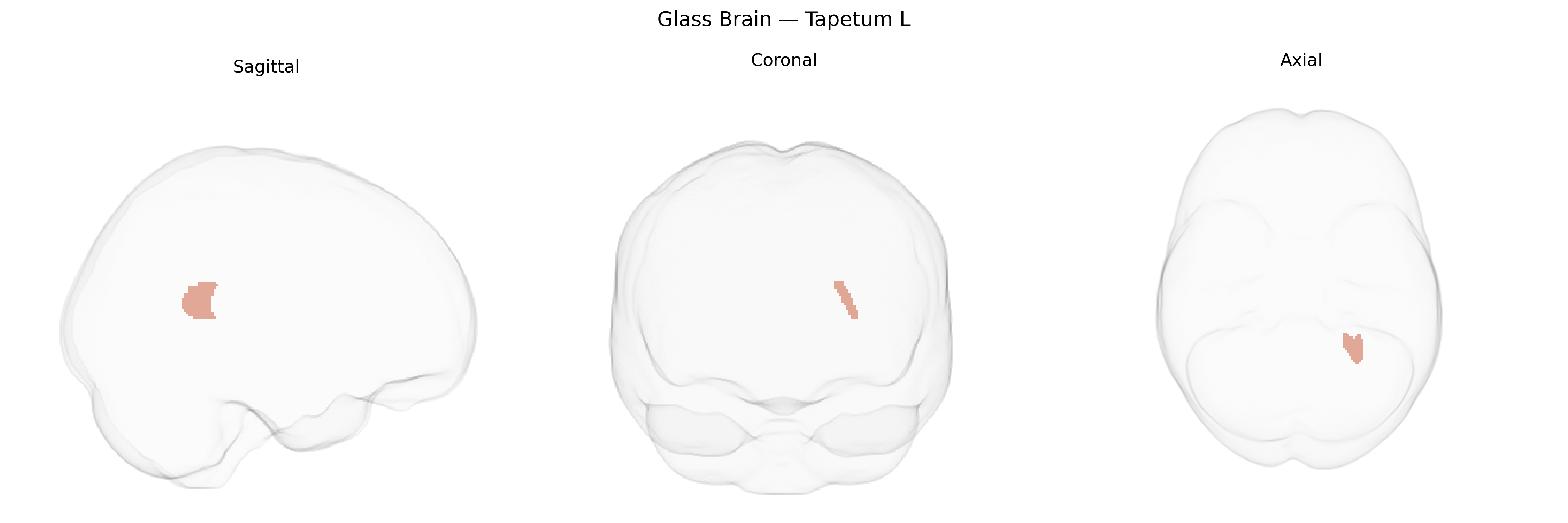
The bilateral Tapetum L in the JHU ICBM labels 1mm Atlas refers to the left tapetum, a thin sheet of commissural fibers within the corpus callosum that runs along the lateral walls of the posterior horn of the lateral ventricle. These myelinated fibers interconnect homologous temporal and occipital cortical regions across hemispheres, contributing to interhemispheric transfer of visual and higher-order associative information. Anatomically, the tapetum is considered part of the corpus callosum’s posterior segment, closely related to the splenium and extending into the temporal lobe white matter. Functionally, its integrity is implicated in coordinated bilateral processing of visual and language-related stimuli, and it is a notable tract in diffusion tensor imaging studies of callosal connectivity. There is no direct Wikipedia article for the tapetum (corpus callosum component); see the related structure Corpus callosum.
The bilateral tapetum of the corpus callosum (L) in the JHU ICBM 1 mm atlas is a small commissural fiber tract connecting temporal lobes, and its microstructural properties (e.g., fractional anisotropy and mean diffusivity) have been examined in large neuroimaging–genetics datasets, though often under broader “corpus callosum” or “temporal callosal fibers” rather than tapetum-specific labels. GWAS of callosal white matter integrity, such as those from ENIGMA and UK Biobank, have identified robust associations between variants in genes involved in axon guidance, myelination, and oligodendrocyte function (including loci near CNTN4, NTRK1/2, and myelin-related genes like MAG and MBP) and diffusion metrics in temporal callosal tracts that overlap the tapetum region, as well as polygenic overlap with educational attainment, intelligence, and psychiatric risk scores (schizophrenia, major depression, bipolar disorder). Tapetum-associated callosal abnormalities have been implicated in disorders with temporal and limbic circuit involvement—most notably schizophrenia, bipolar disorder, and temporal lobe epilepsy—where imaging genetics studies report that risk variants in synaptic and neurodevelopmental genes (for example, CACNA1C, GRIN2A, and ZNF804A) are associated with altered integrity of temporal callosal fibers that include the tapetum. More broadly, corpus callosum tract GWAS demonstrate pleiotropy with neurodevelopmental traits (ADHD, autism spectrum conditions, cognitive performance), suggesting a shared genetic architecture between callosal microstructure and higher-order cognitive and psychiatric phenotypes, but direct tapetum-only GWAS signals remain sparse and are typically inferred from analyses of adjacent or composite callosal and temporal white matter regions rather than isolated tapetum labels.
Overview generated by GPT-4o (2026).
Region ID: 48
Hemisphere: bilateral
Atlas: JHU ICBM labels 1mm
Full Quality Version: Download MP4
Full Quality Version: Download MP4
Wali Sidiqyar*, Gaurav Rudravaram*, Elyssa M. McMaster, Trent M. Schwartz, Adam M. Saunders, Kurt G. Schilling, Bennett A. Landman "Introducing SPINS: A Shared Public Visualization Library of Neuroanatomical Structures." Medical Imaging with Deep Learning- short paper
This resource is licensed under CC0 1.0 Universal (Public Domain).