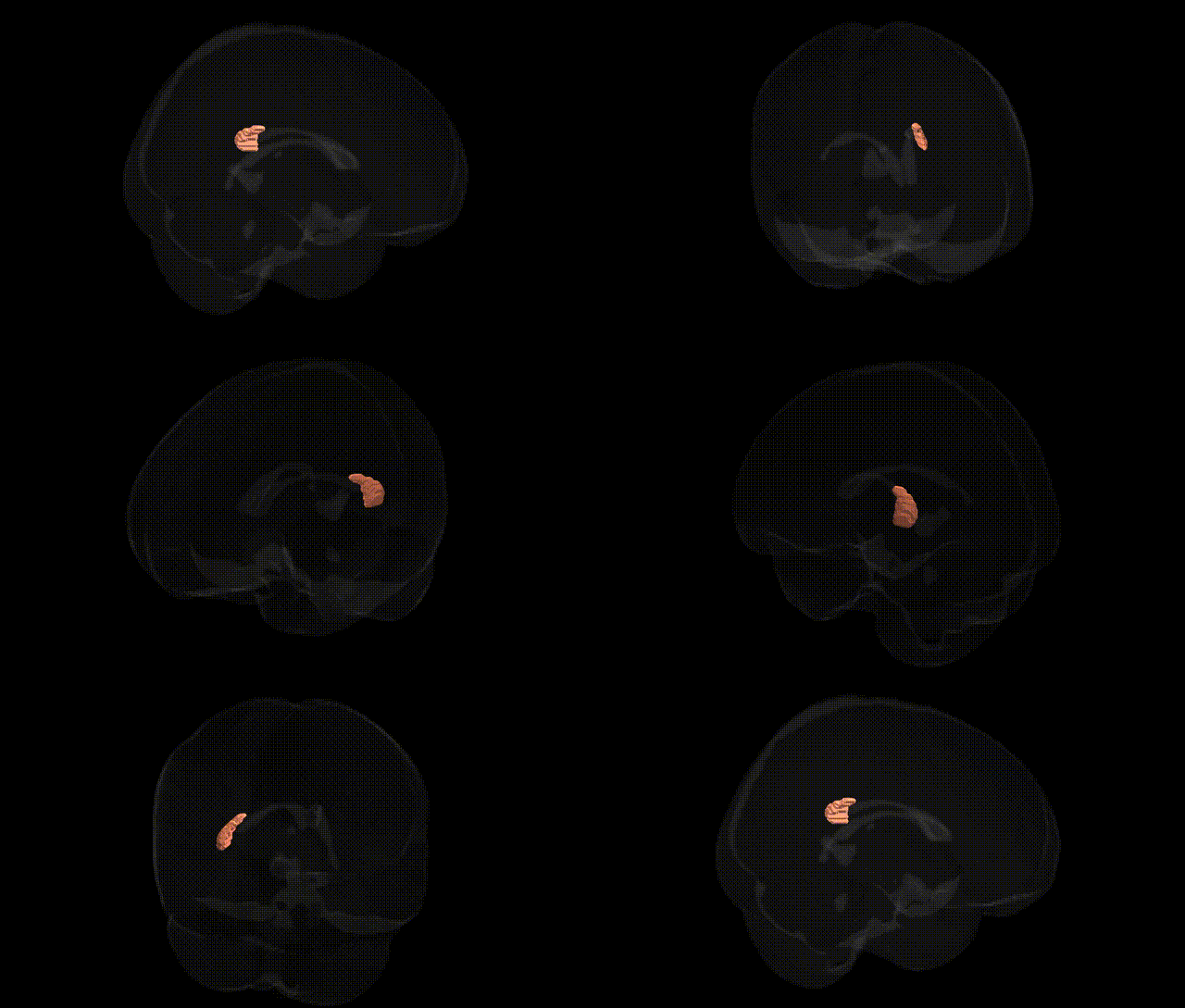
The bilateral tapetum (right) is a white matter structure of the corpus callosum that runs along the lateral ventricles and forms part of the interhemispheric commissural system. In the JHU ICBM 1 mm white matter atlas, the tapetum R label corresponds to callosal fibers coursing in the temporal and occipital regions of the right hemisphere, connecting homologous areas across the midline and contributing to transhemispheric integration of visual and auditory information. These fibers lie adjacent to and partially overlap with other major association and projection pathways but are distinguished by their origin from the splenium of the corpus callosum and their characteristic sheet‑like trajectory along the ventricular wall. There is no direct Wikipedia article for the tapetum of the corpus callosum; a related structure is the Corpus Callosum.
Genetic association studies of white-matter microstructure have implicated the bilateral tapetum—a corpus callosum segment conveying interhemispheric temporal and visual information—as a locus where common variants influence diffusion metrics such as fractional anisotropy and mean diffusivity, although findings are typically reported at the corpus callosum subregion level rather than specifically labeled by the JHU ICBM 1 mm atlas. Large GWAS of diffusion MRI measures (e.g., UK Biobank–based studies) have identified multiple loci in genes involved in axon guidance, myelination, and oligodendrocyte biology (including variants near genes such as NRG1, CNTN4, and MAG) that show significant effects in posterior callosal fibers overlapping the tapetum, suggesting polygenic control of its integrity. Genetically influenced alterations of tapetum or adjacent splenial callosal microstructure have been observed in disorders such as schizophrenia, bipolar disorder, major depression, autism spectrum disorder, and multiple sclerosis, often mediated by risk variants affecting neurodevelopmental and immune pathways that contribute broadly to callosal dysconnectivity. However, no common-variant GWAS signals are known that are uniquely specific to the JHU-defined bilateral tapetum region; instead, associations arise as part of more global callosal or temporal-lobe white-matter phenotypes whose genetic architecture overlaps with that of psychiatric illness, cognitive traits, and neurodegenerative risk.
Overview generated by GPT-4o (2026).
Region ID: 47
Hemisphere: bilateral
Atlas: JHU ICBM labels 1mm
Full Quality Version: Download MP4
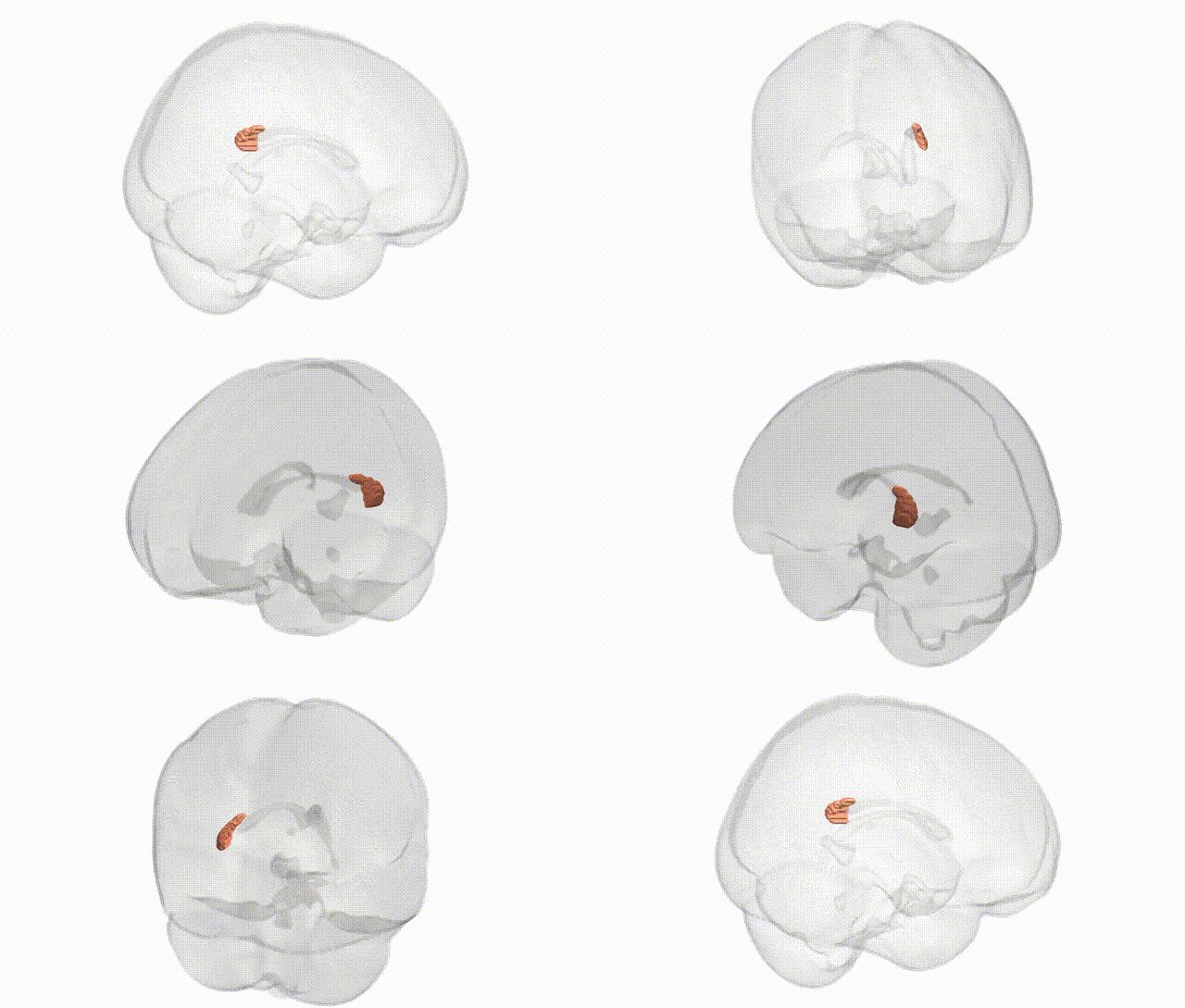
Full Quality Version: Download MP4
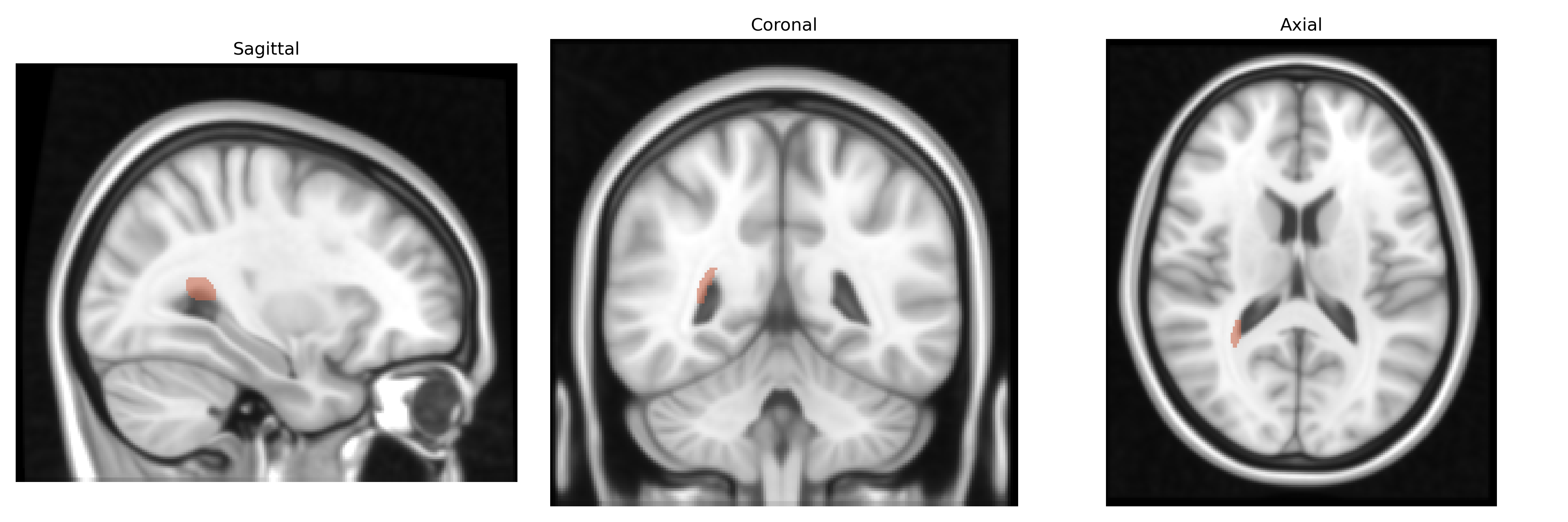
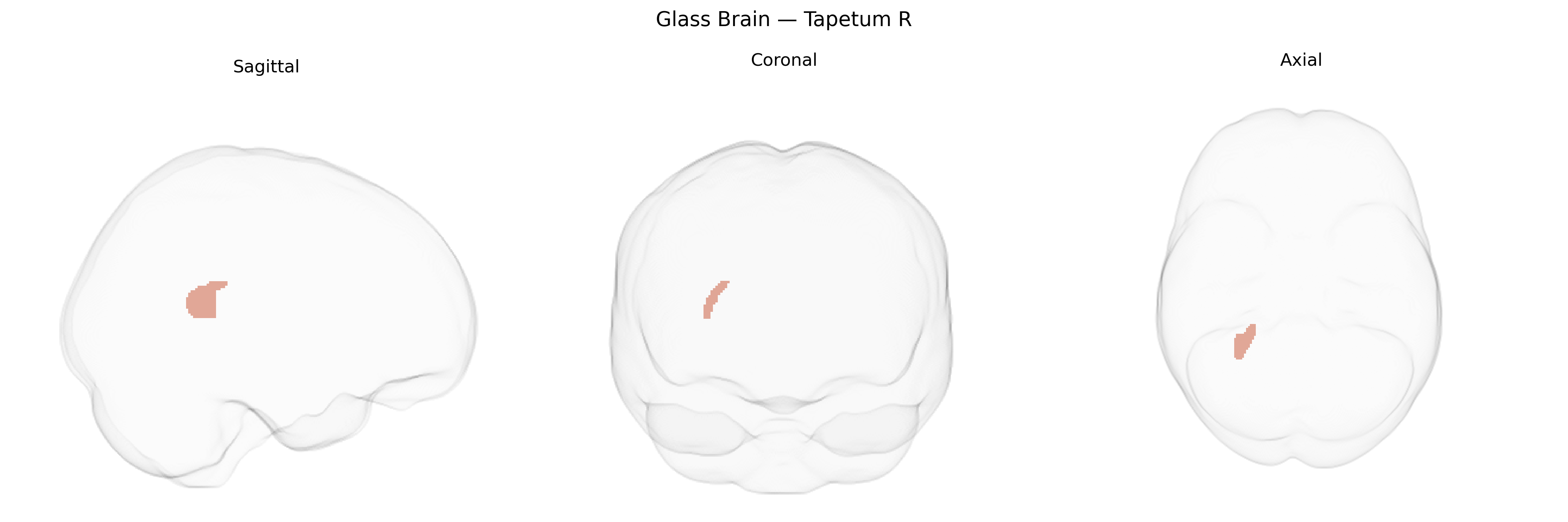
Wali Sidiqyar*, Gaurav Rudravaram*, Elyssa M. McMaster, Trent M. Schwartz, Adam M. Saunders, Kurt G. Schilling, Bennett A. Landman "Introducing SPINS: A Shared Public Visualization Library of Neuroanatomical Structures." Medical Imaging with Deep Learning- short paper
This resource is licensed under CC0 1.0 Universal (Public Domain).