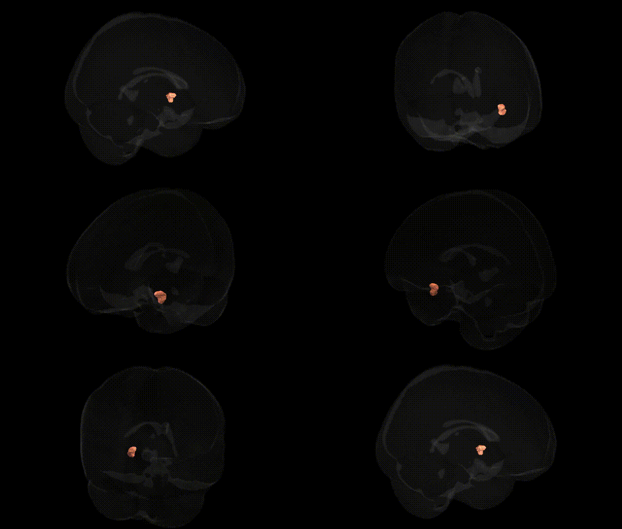
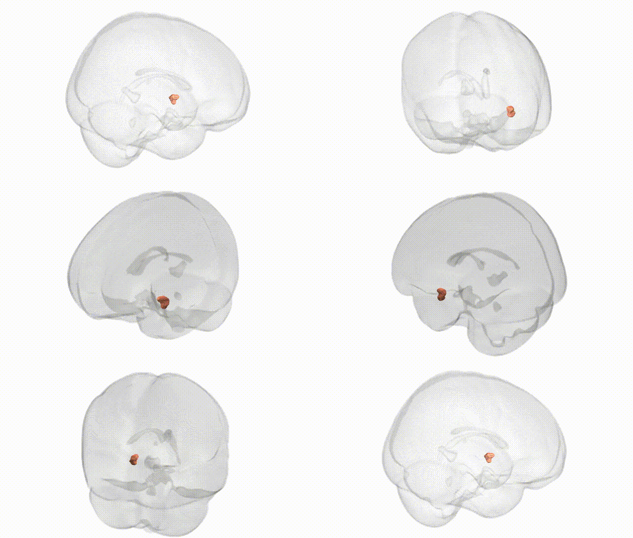
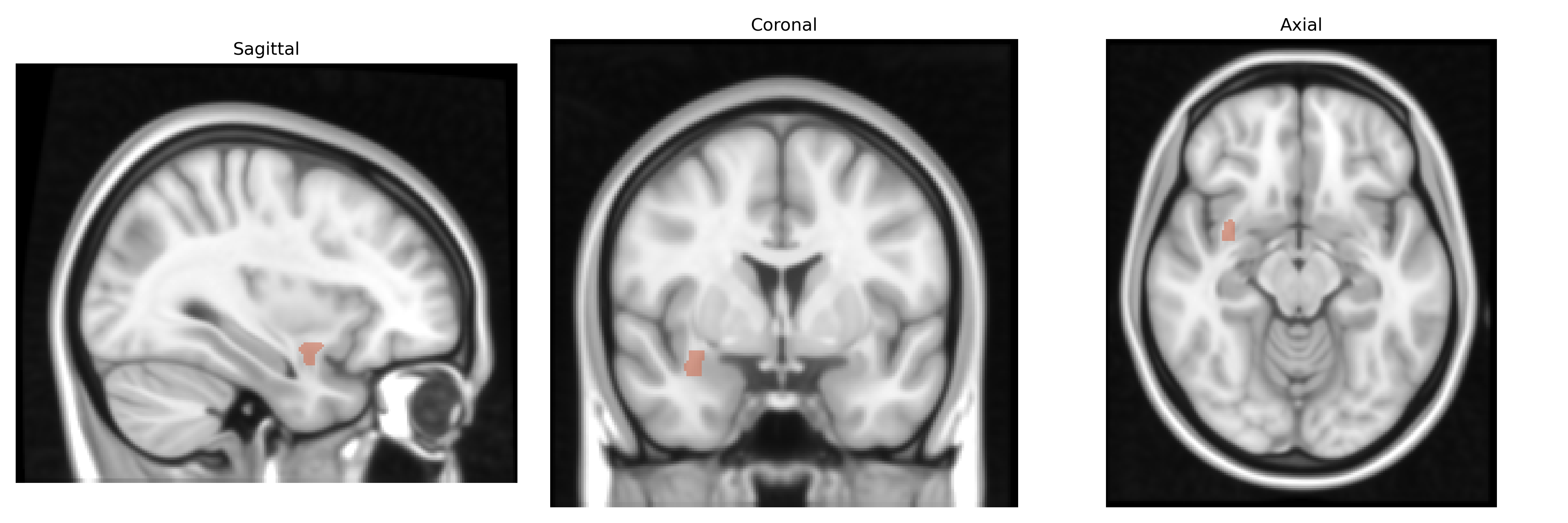
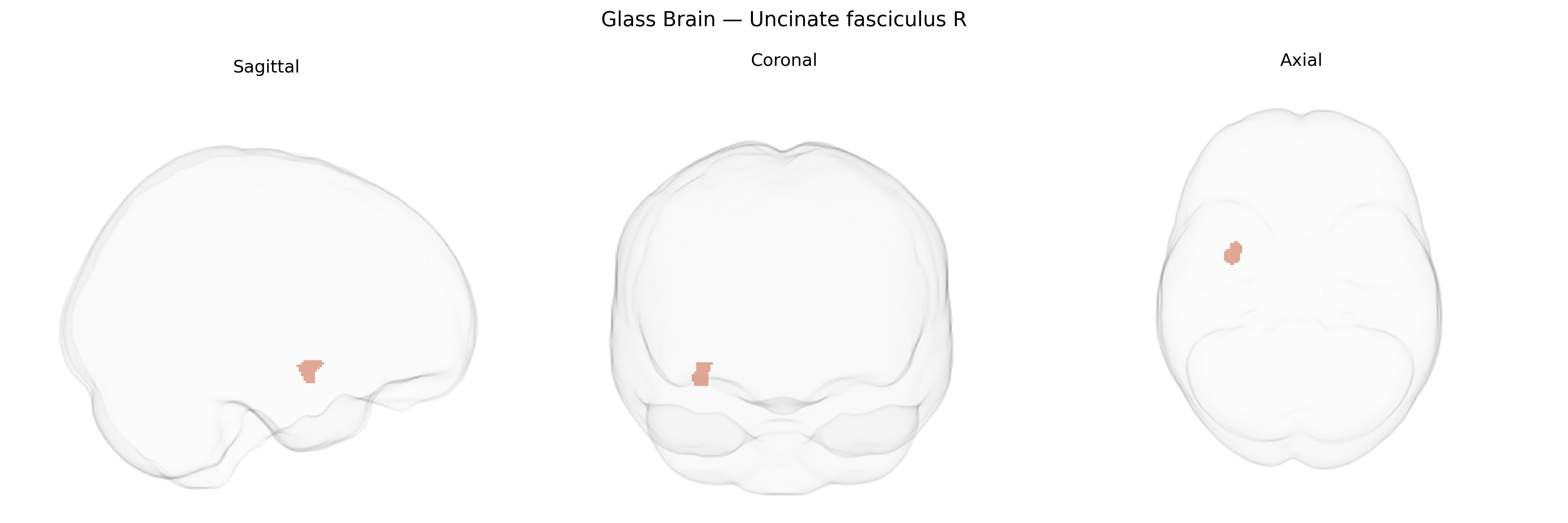
The bilateral Uncinate fasciculus (right) is a long association white matter tract that connects the anterior temporal lobe (including parts of the amygdala and hippocampal region) with the orbitofrontal and ventromedial prefrontal cortices. It courses medially and anteriorly, curving in a hook-like (“uncinate”) shape around the lateral sulcus and through the temporal–frontal junction. Functionally, the uncinate fasciculus is implicated in emotional processing, social cognition, memory retrieval, and aspects of language, integrating limbic information with higher-order executive and decision-making processes in the frontal lobe. In the JHU ICBM labels 1 mm atlas, it is delineated bilaterally as a distinct tract, with the right-side representation often associated with socio-emotional and affective components of behavior. Uncinate fasciculus
The bilateral uncinate fasciculus, as defined in the JHU ICBM 1 mm atlas, has been implicated in multiple genetic and GWAS findings that link structural and microstructural variation in this tract to neuropsychiatric and cognitive phenotypes. Twin and family studies indicate high heritability of uncinate fasciculus fractional anisotropy (FA) and related diffusion metrics, with several large-scale imaging–genetics consortia (e.g., ENIGMA-DTI) identifying common variants in genes involved in myelination, axonal guidance, and neurodevelopment (such as those in the NRG1/ErbB, CNTNAP2, and BDNF pathways) that modulate FA and radial or axial diffusivity in this tract. GWAS of white matter integrity have reported associations between uncinate fasciculus microstructure and loci on chromosomes linked to oligodendrocyte function, lipid metabolism, and neural adhesion molecules, though individual SNP effects are typically small and often fall below genome-wide significance when corrected across tracts. Clinically, genetic risk for schizophrenia, bipolar disorder, major depressive disorder, and autism spectrum disorder—often summarized as polygenic risk scores—has been associated in multiple studies with altered FA or reduced volume in the uncinate fasciculus, consistent with its role in frontotemporal connectivity and emotion regulation. Variants in APOE (particularly ε4) and other Alzheimer’s disease risk genes have been linked to early microstructural changes in the uncinate fasciculus, while GWAS of anxiety traits, neuroticism, and impulsivity have highlighted overlapping polygenic influences on this tract’s integrity. Overall, evidence supports a polygenic architecture in which numerous small-effect variants influencing neurodevelopment, synaptic plasticity, and myelination contribute to inter-individual differences in the uncinate fasciculus and, through this pathway, to vulnerability for mood, psychotic, and neurodegenerative disorders.
Overview generated by GPT-4o (2026).
Region ID: 45
Hemisphere: bilateral
Atlas: JHU ICBM labels 1mm
Full Quality Version: Download MP4
Full Quality Version: Download MP4
Wali Sidiqyar*, Gaurav Rudravaram*, Elyssa M. McMaster, Trent M. Schwartz, Adam M. Saunders, Kurt G. Schilling, Bennett A. Landman "Introducing SPINS: A Shared Public Visualization Library of Neuroanatomical Structures." Medical Imaging with Deep Learning- short paper
This resource is licensed under CC0 1.0 Universal (Public Domain).