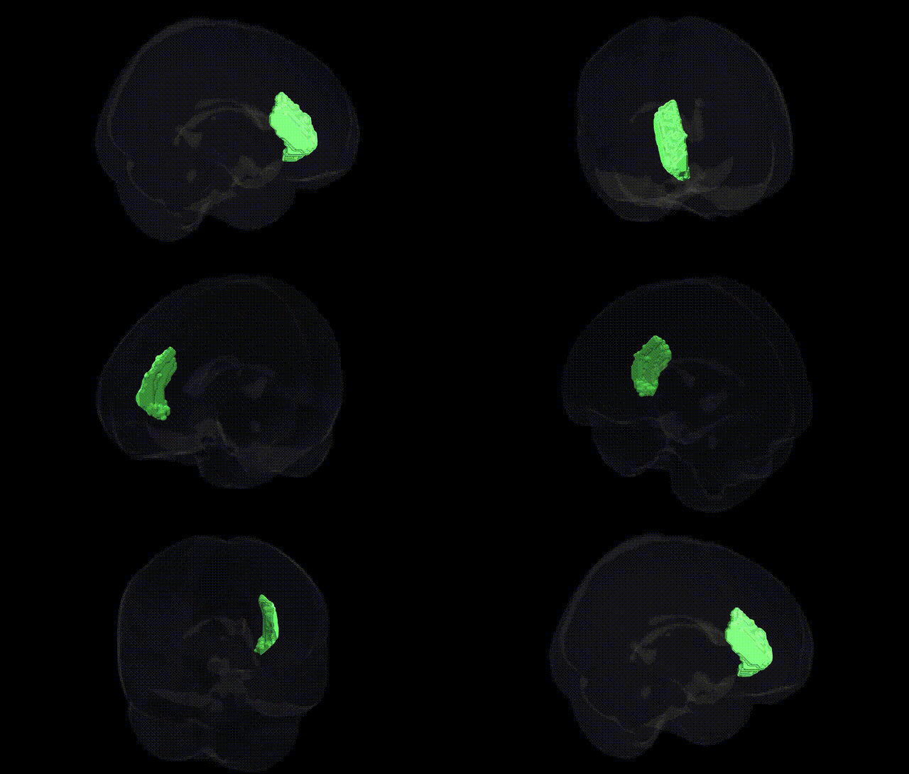
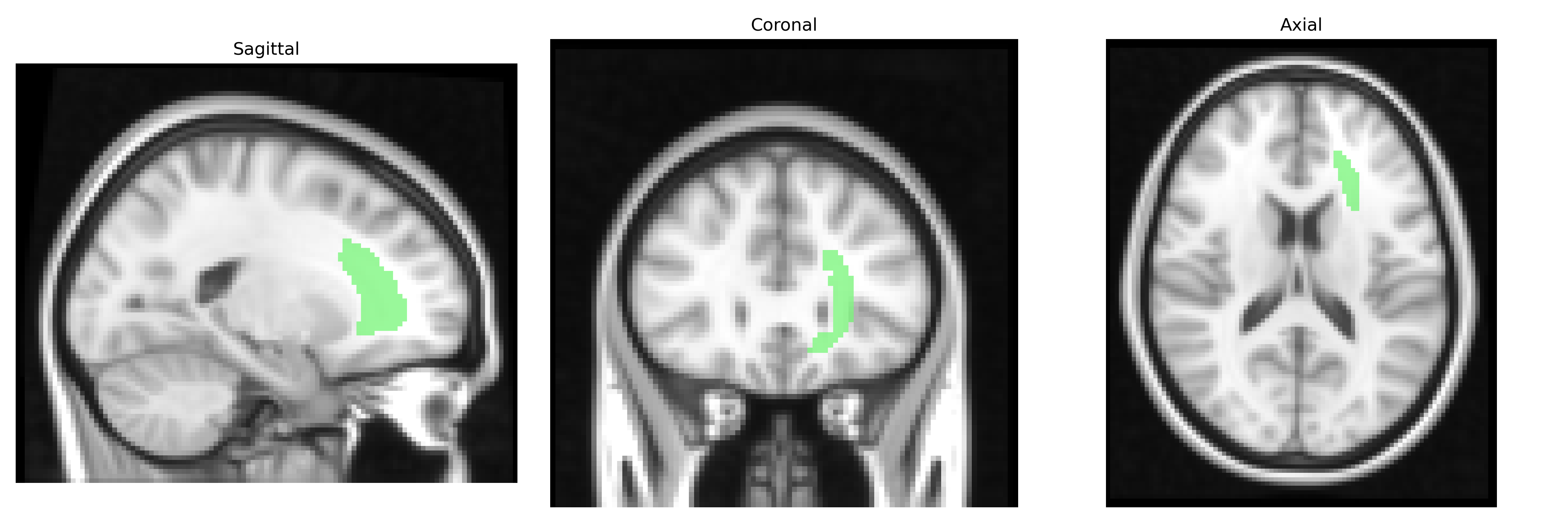
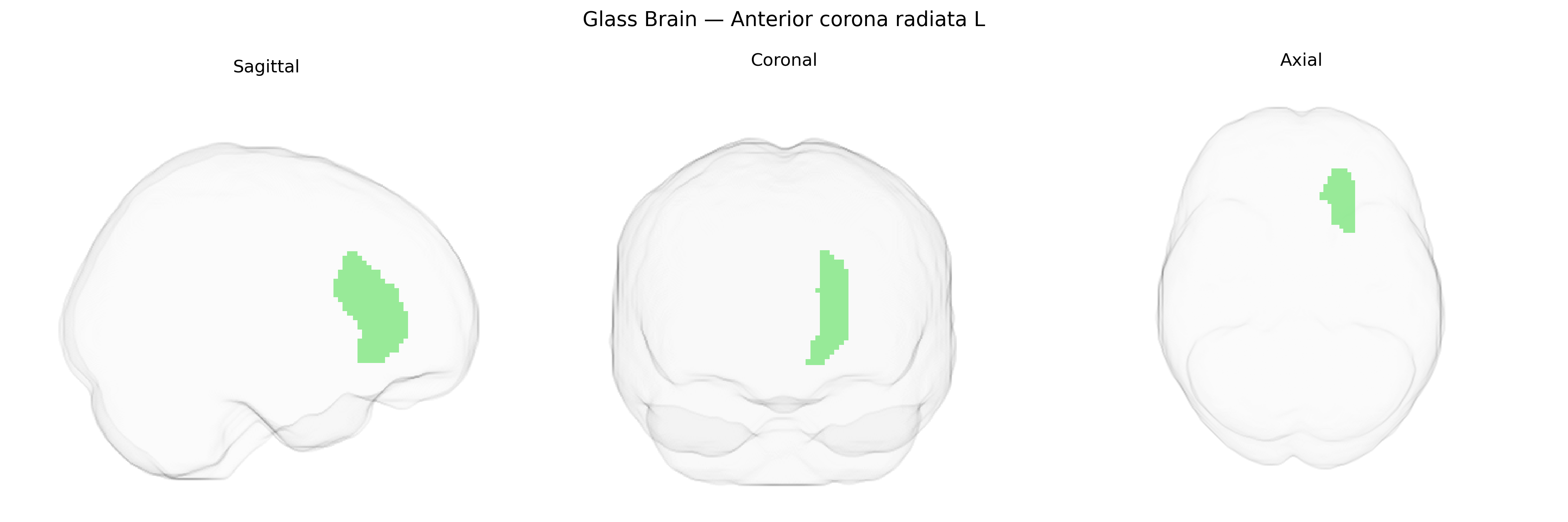
The bilateral anterior corona radiata (L) is a major white matter tract located anterior to the lateral ventricles, forming part of the broader corona radiata that fans out from the internal capsule toward the cerebral cortex. It consists predominantly of ascending and descending myelinated fibers interconnecting frontal cortical regions with subcortical structures, including the thalamus and brainstem, thereby contributing to executive functions, motor planning, attention, and higher-order cognitive processes. In the JHU ICBM 2 mm atlas, this region is defined probabilistically and labeled based on diffusion tensor imaging, serving as a standard reference for structural connectivity and lesion mapping studies in both healthy and clinical populations. There is no direct Wikipedia article for the anterior corona radiata; a related structure encompassing this region is the Corona radiata.
The bilateral anterior corona radiata, a major frontal white-matter pathway labeled in the JHU ICBM 2mm atlas, has shown genetic associations primarily through imaging genetics and GWAS of white-matter microstructure rather than region-specific candidate gene studies. Twin and family-based work indicates substantial heritability of diffusion tensor imaging (DTI) metrics (e.g., fractional anisotropy, mean diffusivity) in this tract, with genome-wide significant loci influencing global or tract-specific white matter that include variants near genes involved in axon guidance, myelination, and neuronal development (such as NTRK1, CNTN4, and NCAM-related pathways), though most signals are not restricted uniquely to the anterior corona radiata. Large-scale GWAS of brain-wide DTI measures have linked variants in or near oligodendrocyte and myelin-related genes (e.g., MAG, PLP1, MBP pathways) to white-matter integrity in frontal tracts that encompass the anterior corona radiata, and polygenic scores for schizophrenia, major depressive disorder, attention-deficit/hyperactivity disorder, and general cognitive ability have been associated with microstructural changes in this region, suggesting that shared genetic architecture contributes to both psychiatric risk and frontal white-matter organization. Additionally, loci related to neurodevelopmental processes and synaptic plasticity identified in GWAS of intelligence, educational attainment, and neuroticism show imaging correlates that include altered anterior corona radiata measures, reinforcing its role as a genetically influenced hub linking cognition, emotion regulation, and vulnerability to mood and psychotic disorders, albeit typically as part of broader fronto-striatal and fronto-limbic networks rather than as an isolated structure.
Overview generated by GPT-4o (2026).
Region ID: 24
Hemisphere: bilateral
Atlas: JHU ICBM labels 2mm
Full Quality Version: Download MP4
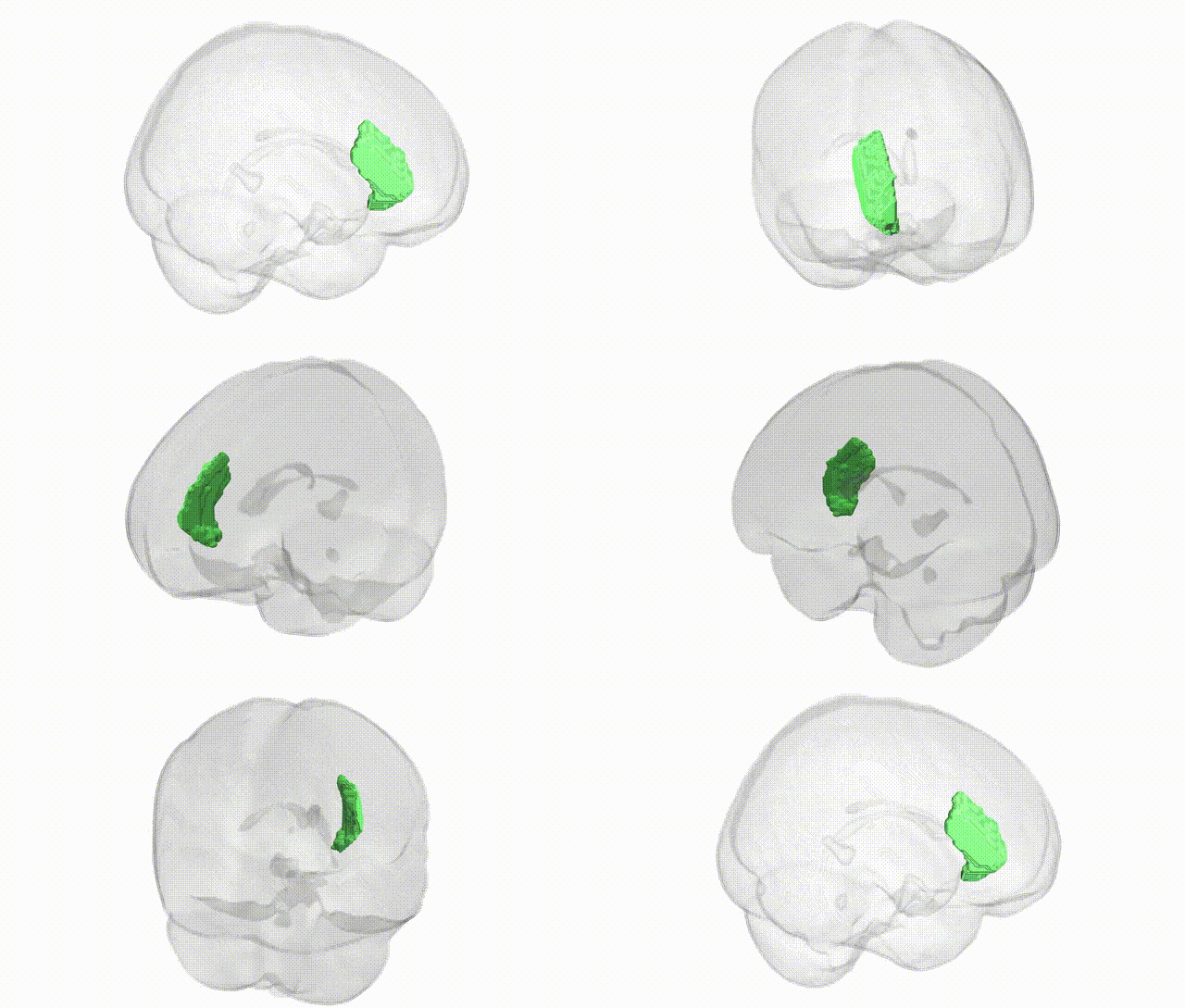
Full Quality Version: Download MP4
Wali Sidiqyar*, Gaurav Rudravaram*, Elyssa M. McMaster, Trent M. Schwartz, Adam M. Saunders, Kurt G. Schilling, Bennett A. Landman "Introducing SPINS: A Shared Public Visualization Library of Neuroanatomical Structures." Medical Imaging with Deep Learning- short paper
This resource is licensed under CC0 1.0 Universal (Public Domain).