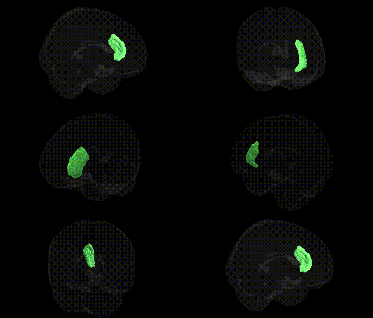
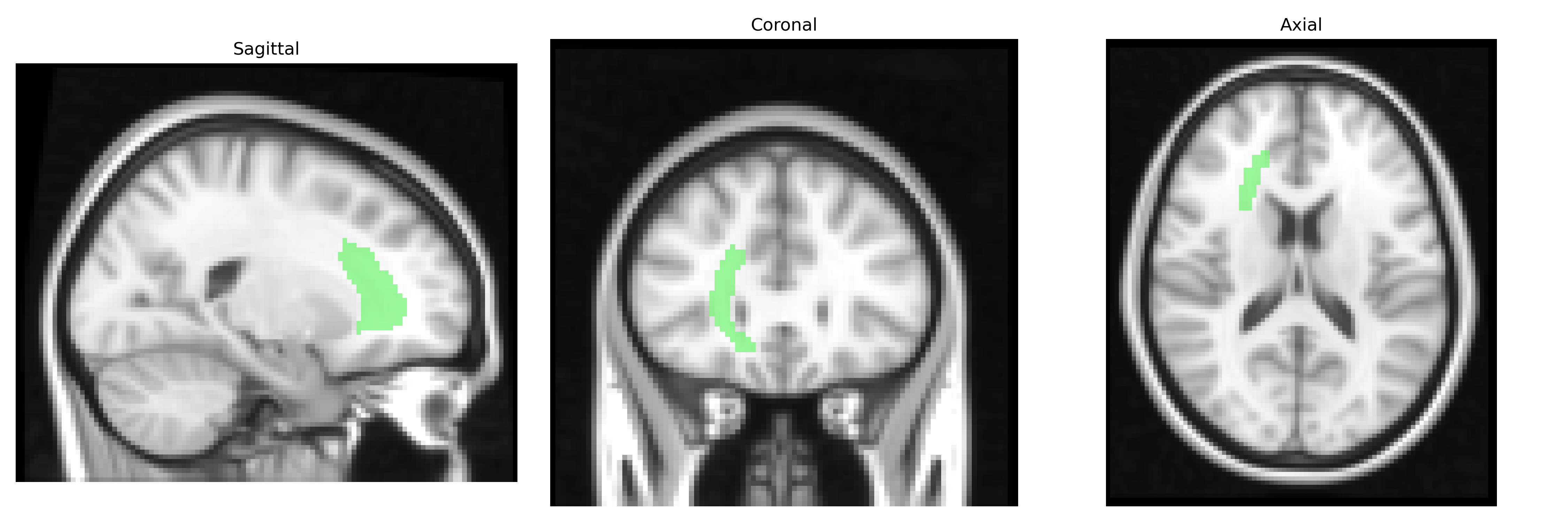
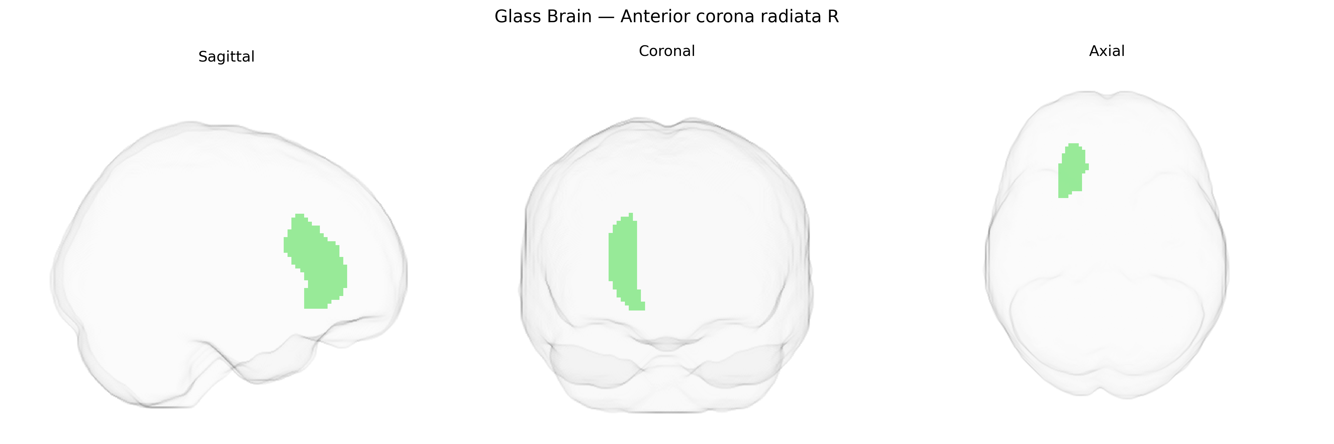
The bilateral Anterior Corona Radiata (Right) is a major white matter tract located in the frontal lobe, composed of ascending and descending projection fibers that fan out from the anterior limb of the internal capsule toward the frontal cortex. It contains fibers connecting the thalamus, brainstem, and subcortical nuclei with prefrontal and premotor regions, and thus participates in higher-order executive functions, attention, working memory, and motor planning. This region is frequently evaluated in diffusion tensor imaging studies due to its vulnerability to microstructural changes in neuropsychiatric and neurodegenerative conditions, as well as in traumatic brain injury and small-vessel cerebrovascular disease. There is no direct Wikipedia article for this specific subregion; a closely related structure is the Corona radiata.
The bilateral anterior corona radiata, as defined in the JHU ICBM labels 2mm Atlas, has been implicated in multiple imaging genetics and GWAS studies through its role as a major frontal white matter pathway connecting prefrontal cortex with subcortical and limbic structures. Variants in genes involved in myelination and axonal integrity—such as NTRK1, CNTN4, MAG, and other neurodevelopmental and cell-adhesion genes—have been associated with diffusion MRI measures (fractional anisotropy, mean diffusivity) in this tract, often in large consortia-based GWAS of white matter microstructure. Altered genetic influences on the anterior corona radiata have been reported in relation to schizophrenia, bipolar disorder, major depression, ADHD, and autism spectrum conditions, where risk variants in synaptic and neurodevelopmental genes show downstream effects on frontal white matter integrity. Polygenic risk scores for psychiatric disorders and cognitive traits, including intelligence and educational attainment, have also been linked to structural variation in the anterior corona radiata, suggesting that distributed genetic architecture governing cortical development, oligodendrocyte function, and synaptic plasticity contributes to inter-individual differences in this region. Additionally, GWAS of brain connectivity and white matter hyperintensities in aging and small vessel disease have identified loci influencing anterior corona radiata structure, supporting its role as a genetically modulated substrate for both neuropsychiatric vulnerability and vascular-related cognitive decline.
Overview generated by GPT-4o (2026).
Region ID: 23
Hemisphere: bilateral
Atlas: JHU ICBM labels 2mm
Full Quality Version: Download MP4
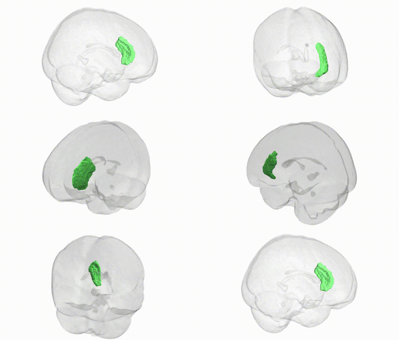
Full Quality Version: Download MP4
Wali Sidiqyar*, Gaurav Rudravaram*, Elyssa M. McMaster, Trent M. Schwartz, Adam M. Saunders, Kurt G. Schilling, Bennett A. Landman "Introducing SPINS: A Shared Public Visualization Library of Neuroanatomical Structures." Medical Imaging with Deep Learning- short paper
This resource is licensed under CC0 1.0 Universal (Public Domain).