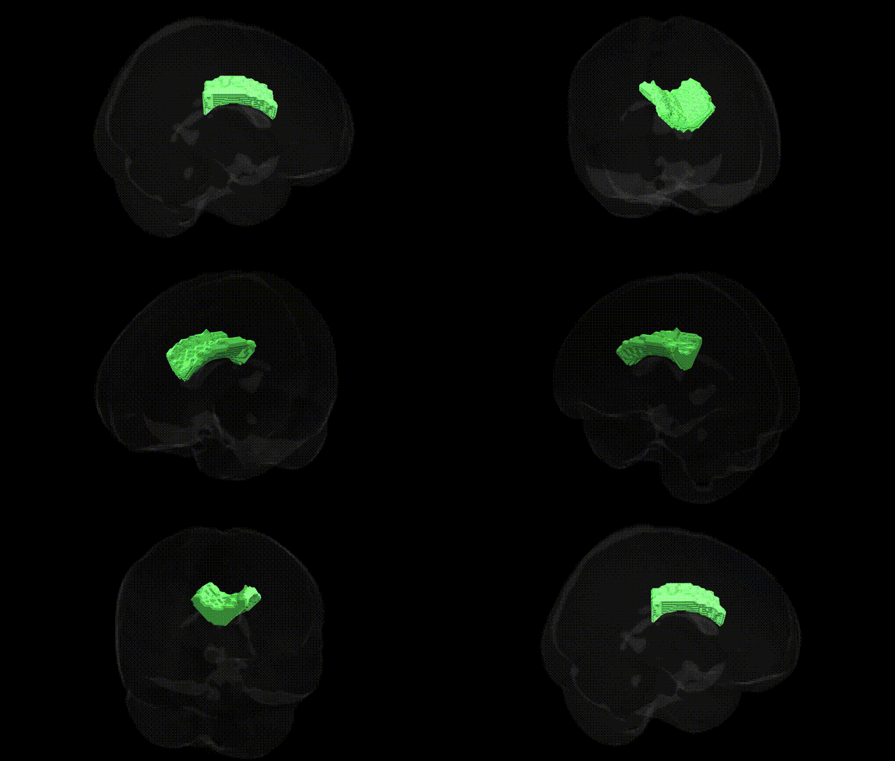
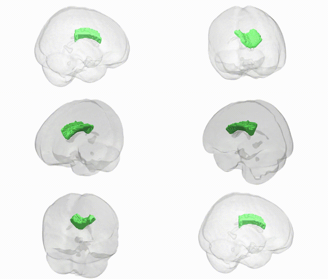
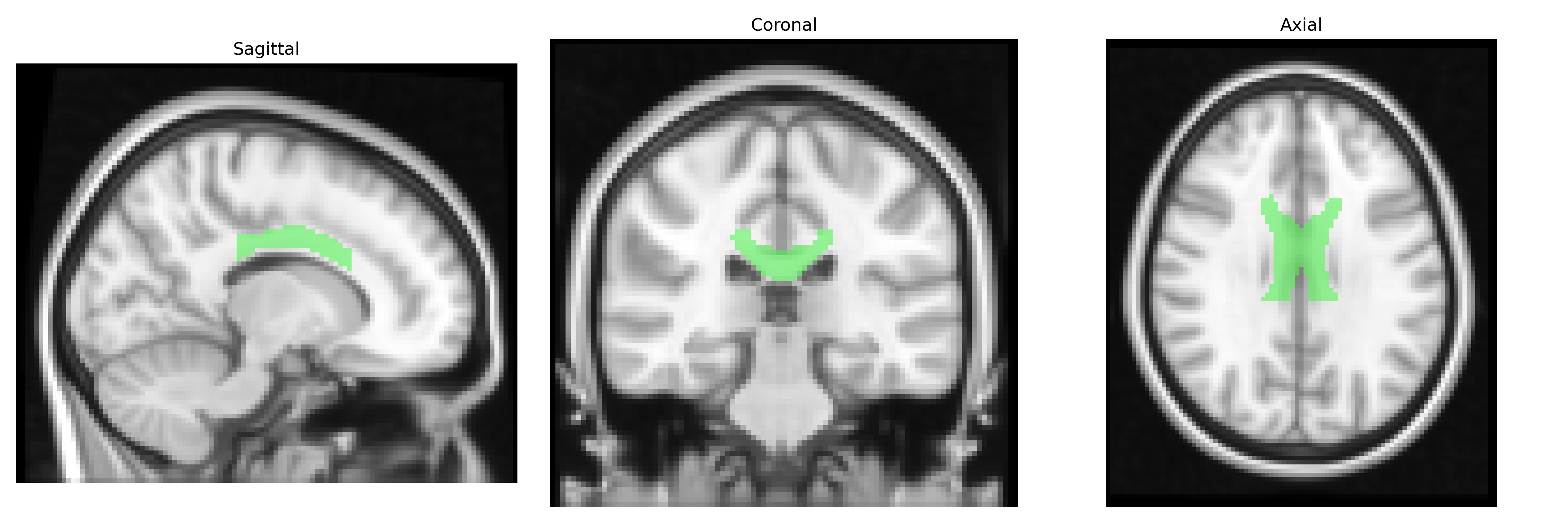
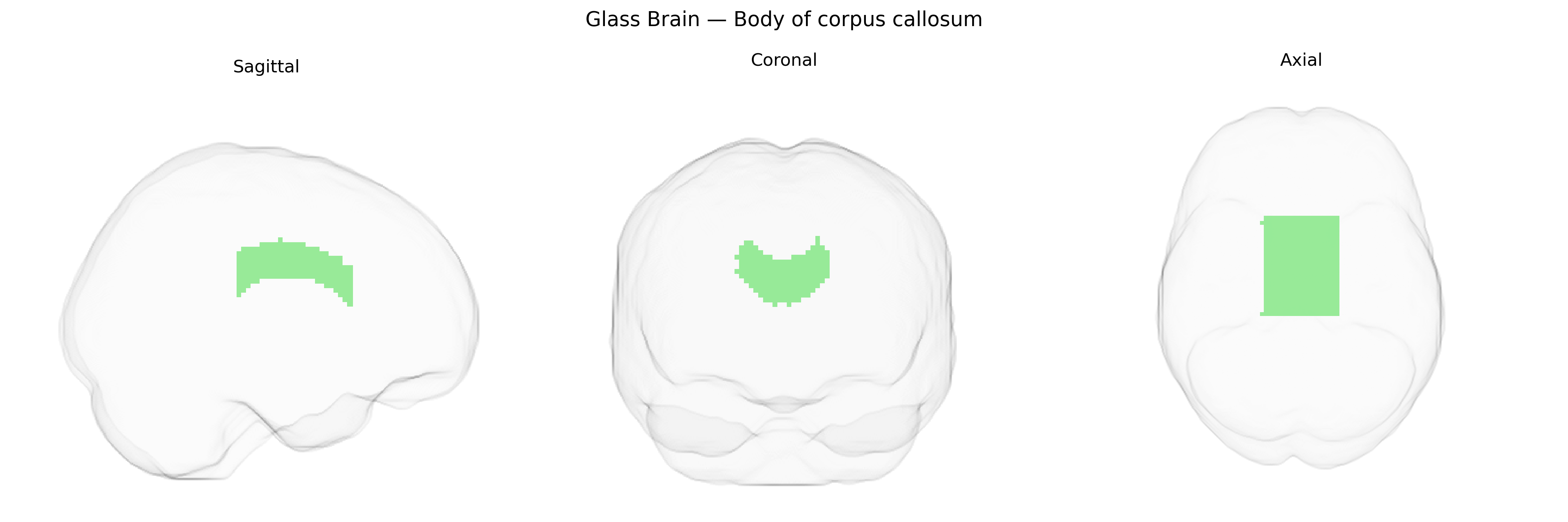
The bilateral Body of corpus callosum, as defined in the JHU ICBM 2 mm atlas, corresponds to the central segment of the corpus callosum, a major commissural white matter tract that interconnects homologous cortical areas of both cerebral hemispheres. Composed primarily of densely packed myelinated axons, this region facilitates interhemispheric transfer of sensory, motor, and associative information, especially between parietal and posterior frontal cortices. It plays a critical role in coordinated bimanual movements, integration of higher-order cognitive processes, and maintenance of unified perception. Damage or agenesis in this area can result in disconnection syndromes, including deficits in interhemispheric communication and higher-order cognitive impairment. Corpus callosum
The bilateral body of the corpus callosum, as defined in the JHU ICBM 2mm atlas, has been implicated in numerous imaging-genetics and GWAS-based endophenotype studies, showing heritable variation in callosal volume, thickness, and microstructural metrics (e.g., fractional anisotropy and mean diffusivity) linked to polygenic influences on neurodevelopment and myelination. Large-scale neuroimaging GWAS (such as ENIGMA and UK Biobank–based analyses) have identified common variants near or within genes involved in axon guidance (e.g., ROBO1/ROBO2, DCC, NTN1), cell adhesion and midline crossing (e.g., L1CAM-related pathways), and oligodendrocyte function/myelin biology (e.g., MAG, MBP, and other myelin-related loci) that modulate callosal structure, including the callosal body. Clinically, rare pathogenic mutations in genes such as L1CAM, ARX, and others cause partial or complete agenesis or dysgenesis of the corpus callosum, often involving the callosal body, in neurodevelopmental disorders including intellectual disability, epilepsy, and syndromic forms of autism. Imaging-genetic work also links callosal body alterations to polygenic risk for schizophrenia, bipolar disorder, major depression, and ADHD, with associated loci often pointing to synaptic, neurodevelopmental, and myelination pathways. Additionally, genetic variants associated with neurodegenerative and neuroinflammatory conditions (e.g., multiple sclerosis risk loci influencing demyelination) have been associated with microstructural changes in the callosal body, supporting its role as a convergent anatomical target of diverse genetic risks affecting interhemispheric connectivity.
Overview generated by GPT-4o (2026).
Region ID: 4
Hemisphere: bilateral
Atlas: JHU ICBM labels 2mm
Full Quality Version: Download MP4
Full Quality Version: Download MP4
Wali Sidiqyar*, Gaurav Rudravaram*, Elyssa M. McMaster, Trent M. Schwartz, Adam M. Saunders, Kurt G. Schilling, Bennett A. Landman "Introducing SPINS: A Shared Public Visualization Library of Neuroanatomical Structures." Medical Imaging with Deep Learning- short paper
This resource is licensed under CC0 1.0 Universal (Public Domain).