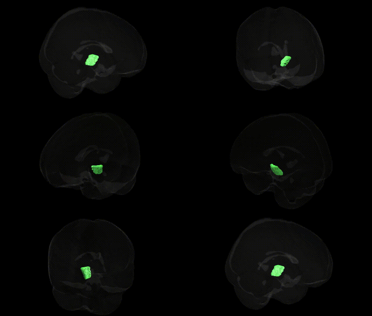
The bilateral cerebral peduncles (right side represented in the JHU ICBM labels 2 mm atlas) are paired bundles of white matter located in the ventral midbrain, forming the most anterior portion of the midbrain and continuous rostrally with the internal capsule and caudally with the pons. They consist predominantly of descending corticospinal, corticobulbar, and corticopontine fibers that transmit motor commands from the cerebral cortex to the brainstem and spinal cord, playing a key role in voluntary movement control and motor coordination. These structures are organized somatotopically, with different fiber tracts occupying distinct medial–lateral positions, and are closely associated with neighboring midbrain structures such as the substantia nigra and tegmentum. Lesions in the cerebral peduncles can result in characteristic motor deficits, including contralateral weakness and movement disorders, reflecting disruption of major descending motor pathways. Cerebral peduncle
The bilateral cerebral peduncle (right) as defined in the JHU ICBM 2 mm atlas is a major white matter pathway containing descending corticospinal, corticobulbar, and corticopontine fibers, and while few genome-wide association studies (GWAS) focus on this specific labeled region by name, several large neuroimaging genetics consortia (e.g., ENIGMA, UK Biobank) have reported heritable variation and genetic influences on microstructural measures such as fractional anisotropy (FA) and mean diffusivity (MD) within the cerebral peduncles. Common variants in genes involved in axon guidance, myelination, and oligodendrocyte function (e.g., CNTN4, NTRK3, MAG, MBP) have been implicated in global and tract-specific white matter integrity, occasionally including brainstem and peduncular tracts in voxelwise or tract-based spatial statistics analyses, although such findings are usually reported at the level of broader white matter networks rather than the cerebral peduncle alone. Genetic risk for neurodegenerative and neurodevelopmental disorders that affect corticospinal pathways—such as amyotrophic lateral sclerosis (ALS), hereditary spastic paraplegia, and certain forms of multiple sclerosis—has been associated with structural and diffusion changes in the cerebral peduncles, but the implicated loci (e.g., C9orf72, SOD1, SPAST, various HLA and immune-related variants) are linked to disease processes rather than selectively to this tract. In psychiatric and cognitive GWAS, polygenic scores for schizophrenia, bipolar disorder, major depression, and general cognitive ability have been associated with global white matter microstructure and brainstem connectivity, sometimes including the cerebral peduncular region, yet these effects are diffuse and not specific to the right cerebral peduncle. Overall, current genetic evidence supports substantial heritability of diffusion MRI metrics within the cerebral peduncles and implicates genes involved in neurodevelopment, myelination, and neurodegeneration, but no robust, widely replicated GWAS signal is uniquely or specifically tied to the JHU-defined bilateral cerebral peduncle R region as an isolated locus.
Overview generated by GPT-4o (2026).
Region ID: 15
Hemisphere: bilateral
Atlas: JHU ICBM labels 2mm
Full Quality Version: Download MP4
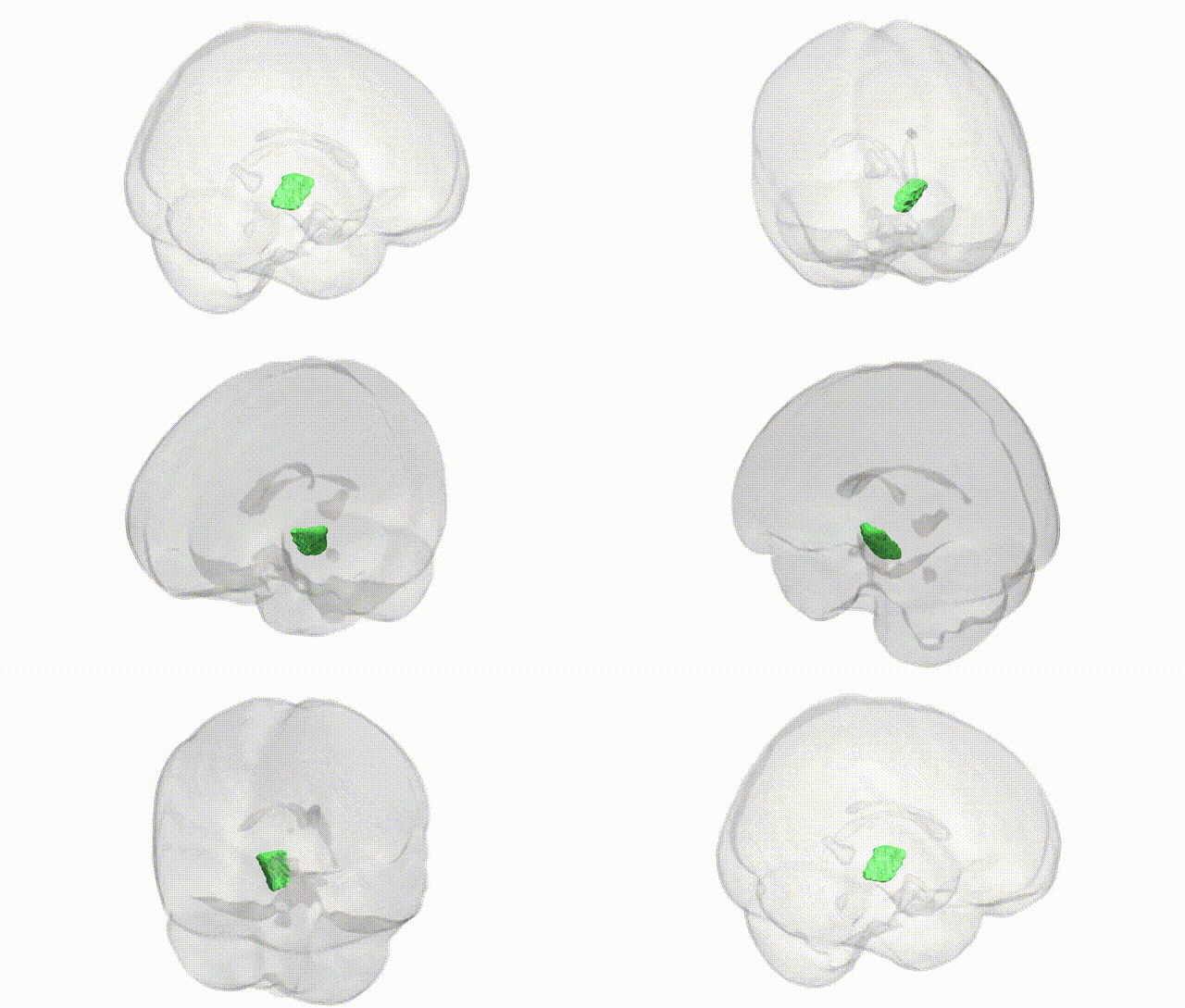
Full Quality Version: Download MP4
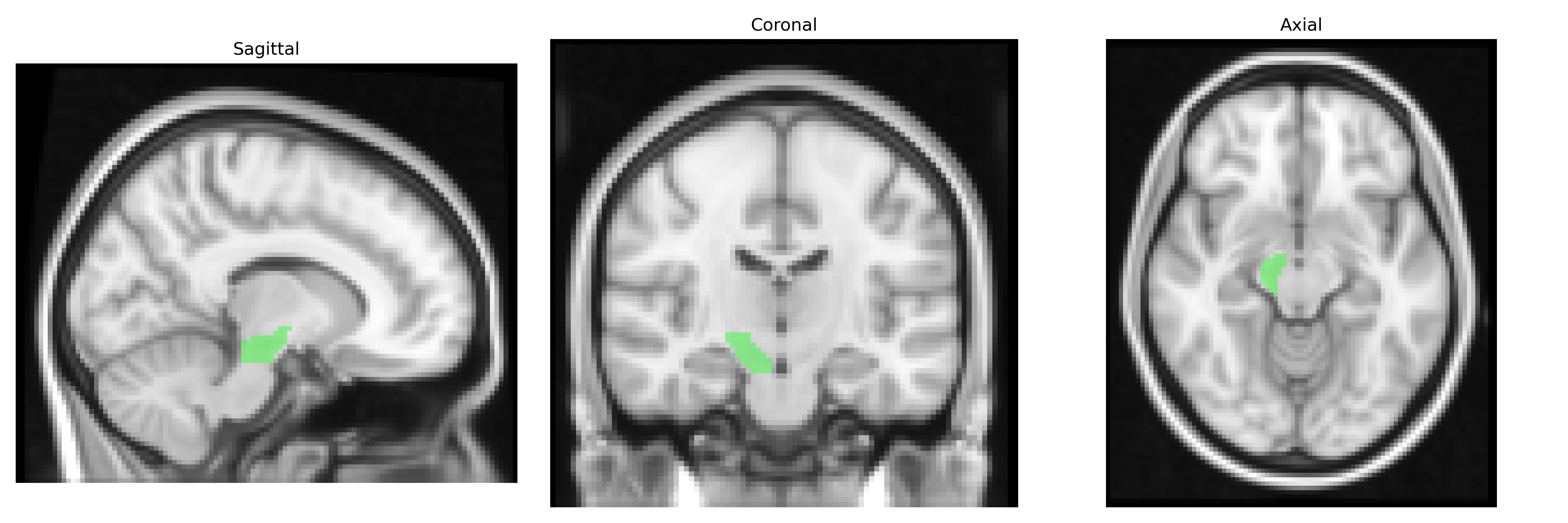
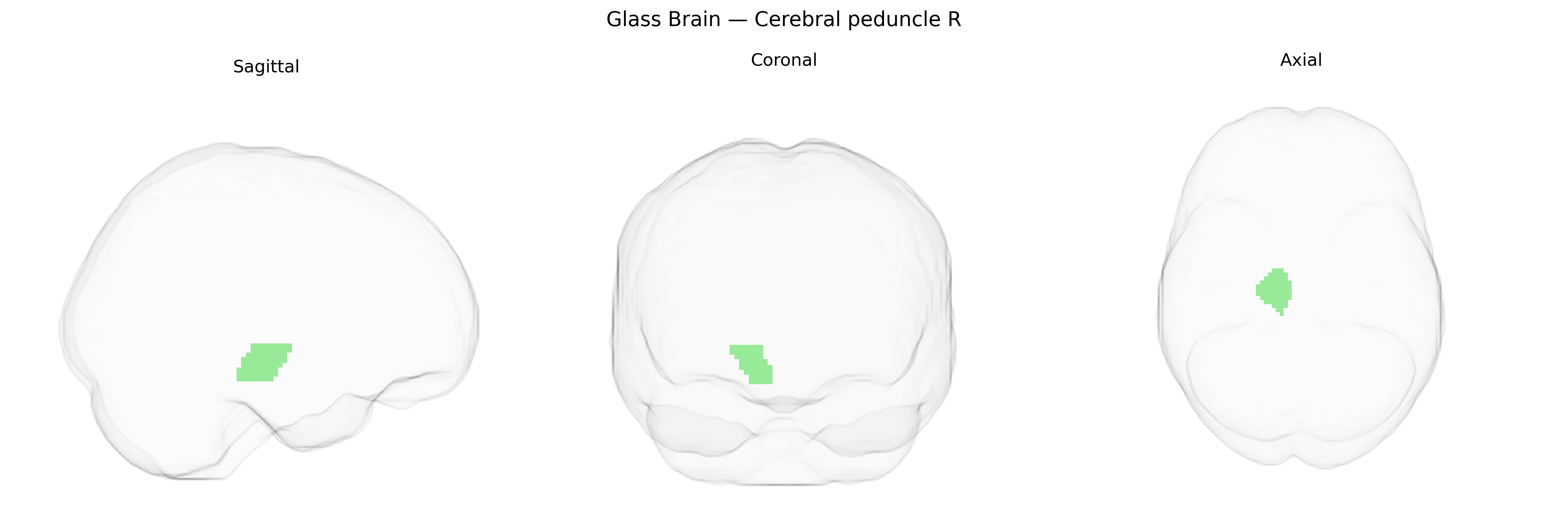
Wali Sidiqyar*, Gaurav Rudravaram*, Elyssa M. McMaster, Trent M. Schwartz, Adam M. Saunders, Kurt G. Schilling, Bennett A. Landman "Introducing SPINS: A Shared Public Visualization Library of Neuroanatomical Structures." Medical Imaging with Deep Learning- short paper
This resource is licensed under CC0 1.0 Universal (Public Domain).