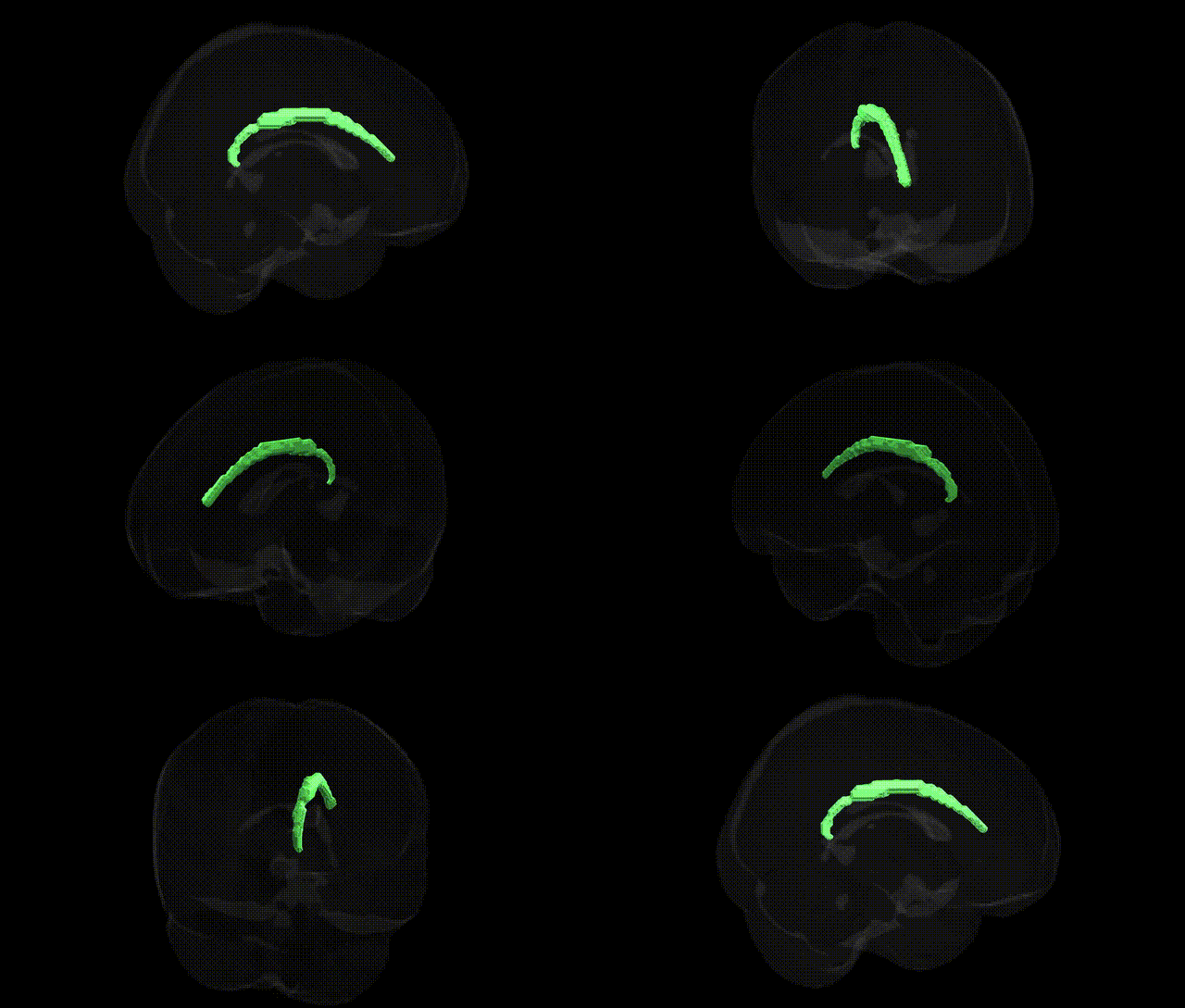
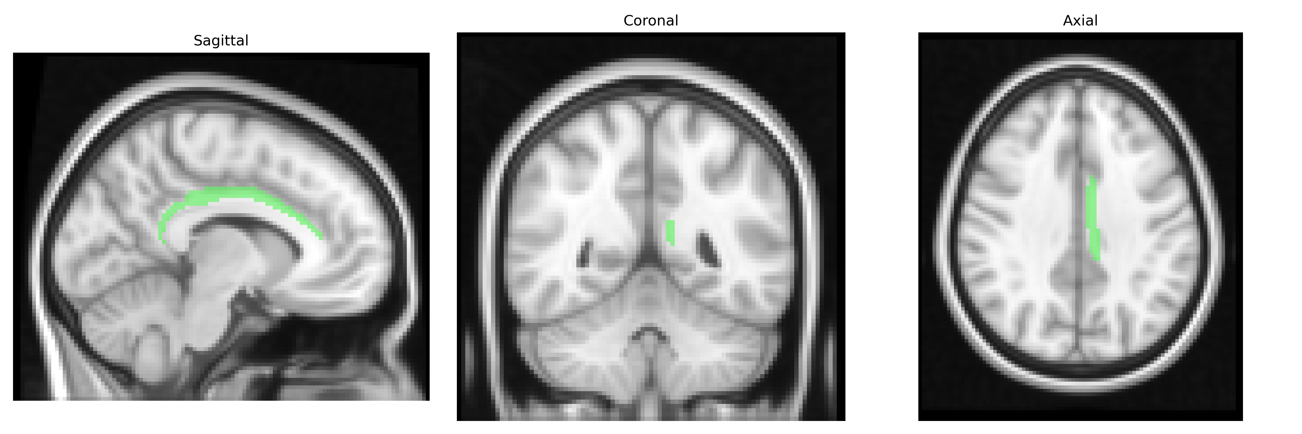
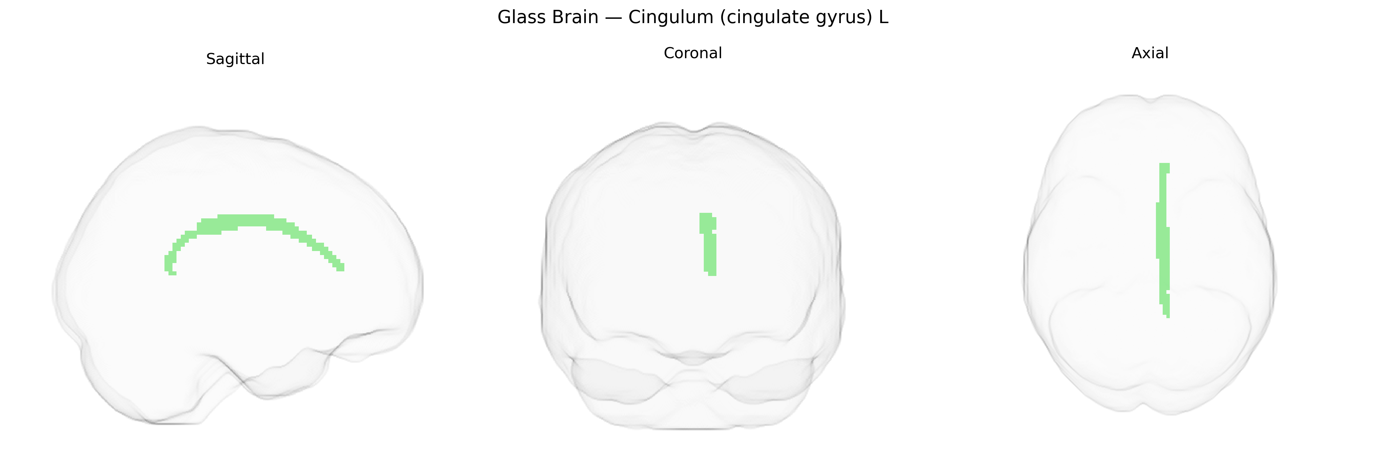
The bilateral Cingulum (cingulate gyrus) L, as defined in the JHU ICBM DTI-based white matter atlas, corresponds to the left-hemisphere segment of a major long association fiber bundle coursing within the cingulate gyrus and extending around the corpus callosum. This tract interconnects medial frontal, parietal, and temporal regions, forming a critical component of the limbic system involved in higher-order cognitive integration, emotional regulation, and memory processes, particularly episodic and autobiographical memory. Structurally, the cingulum lies in the white matter beneath the cingulate cortex, following a curved trajectory that links the anterior cingulate and prefrontal areas with posterior cingulate and retrosplenial cortices, and further toward parahippocampal regions, thereby supporting communication among networks subserving attention, internal mentation, and affective processing. There is no direct Wikipedia article for this tract; see the related cortical region Cingulate gyrus.
The bilateral cingulum (cingulate gyrus) as defined in the JHU ICBM 2 mm atlas has been repeatedly implicated in imaging genetics and GWAS of brain structure and connectivity, with common variants influencing its microstructure (e.g., fractional anisotropy) and volume. Large neuroimaging GWAS consortia such as ENIGMA and UK Biobank have identified associations between cingulum white matter integrity and loci in or near genes involved in axon guidance, myelination, and synaptic function, including but not limited to variants near CNTNAP2, NRG1, and genes in the MAPT region, although specific signals often fall in noncoding regulatory regions. Cingulum-related measures have been genetically correlated with cognitive performance, educational attainment, and neuroticism, and polygenic scores for these traits predict variation in cingulum structure. In clinical imaging-genetic studies, altered cingulum integrity has been linked to schizophrenia, major depressive disorder, bipolar disorder, obsessive–compulsive disorder, PTSD, and Alzheimer’s disease, with some evidence that risk alleles for these disorders (for example in CACNA1C, ZNF804A, and APOE) modulate cingulum connectivity or microstructure. Additionally, GWAS of chronic pain, anxiety, and internalizing symptoms report shared genetic architecture with cingulate and cingulum metrics, consistent with the role of this tract in emotion regulation and pain processing, though most associations currently identify distributed polygenic influences rather than strong region-specific effects.
Overview generated by GPT-4o (2026).
Region ID: 36
Hemisphere: bilateral
Atlas: JHU ICBM labels 2mm
Full Quality Version: Download MP4
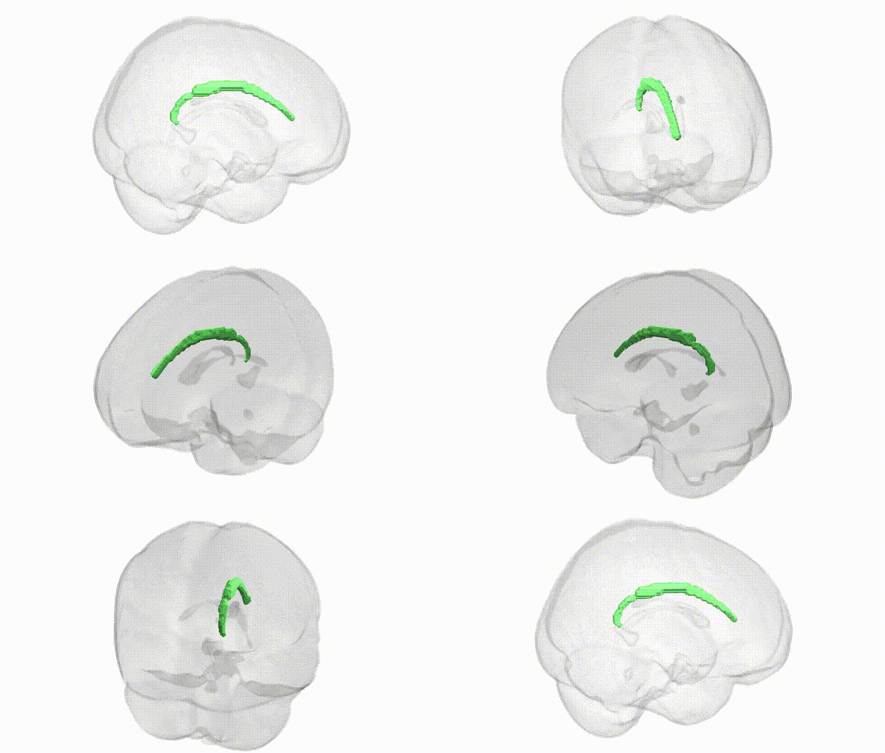
Full Quality Version: Download MP4
Wali Sidiqyar*, Gaurav Rudravaram*, Elyssa M. McMaster, Trent M. Schwartz, Adam M. Saunders, Kurt G. Schilling, Bennett A. Landman "Introducing SPINS: A Shared Public Visualization Library of Neuroanatomical Structures." Medical Imaging with Deep Learning- short paper
This resource is licensed under CC0 1.0 Universal (Public Domain).