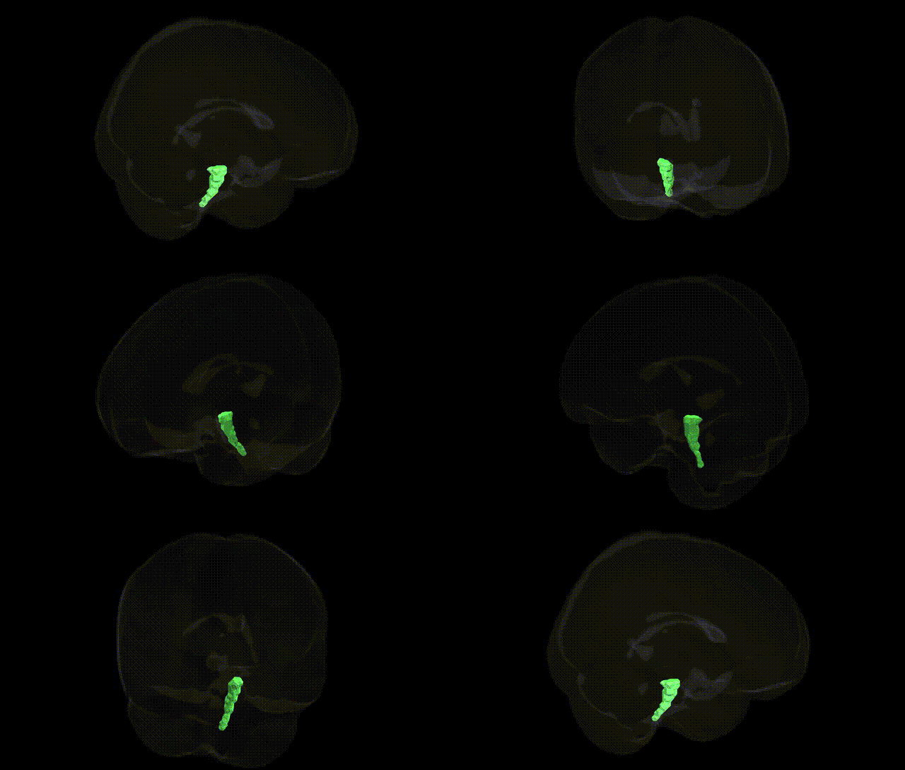
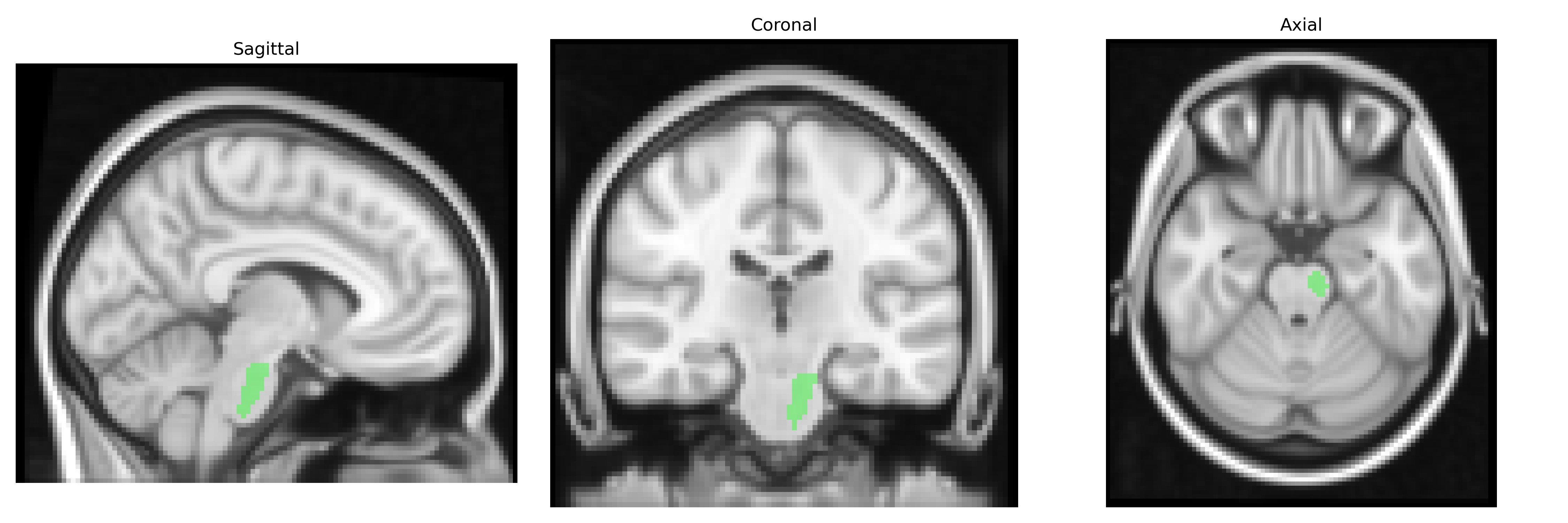
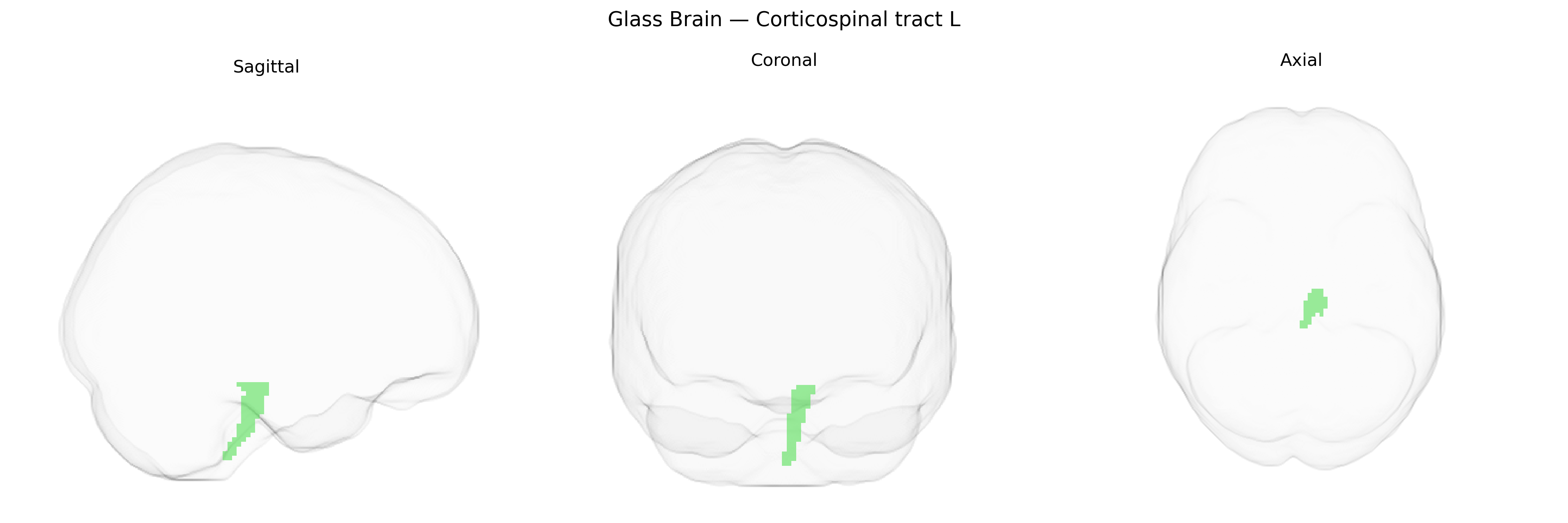
The bilateral corticospinal tract (L) in the JHU ICBM 2mm atlas refers to the left-sided portion of the major descending motor pathway that transmits signals from the cerebral cortex to the spinal cord, primarily originating from neurons in the primary motor cortex, premotor cortex, and supplementary motor areas. Fibers descend through the corona radiata and internal capsule, traverse the cerebral peduncles and brainstem, and largely decussate at the pyramidal decussation in the caudal medulla before continuing in the lateral funiculus of the spinal cord to terminate on interneurons and α-motor neurons. Functionally, this tract is critical for voluntary, fine, and fractionated movements of the distal limbs, especially precise hand and finger control, and lesions affecting the left corticospinal tract typically result in contralateral motor deficits such as weakness, spasticity, and loss of skilled movement. There is no dedicated Wikipedia article for the “corticospinal tract (L)” as an atlas label; a related and encompassing structure is the Corticospinal tract.
The bilateral corticospinal tract (CST) in the left hemisphere, as defined in the JHU ICBM 2 mm atlas, has been implicated in multiple imaging-genetics and GWAS-based endophenotype studies, which generally examine CST microstructure (e.g., fractional anisotropy) as a heritable trait rather than linking genes to the atlas label per se. Twin and family studies show high heritability of CST integrity, with variants in genes involved in axon guidance, myelination, and neuronal plasticity (such as NTRK1/2, CNTN4, MAG, and PLP1) repeatedly associated with diffusion metrics in CST and other major white-matter tracts. Large-scale brain-structure GWAS (e.g., ENIGMA and UK Biobank–based analyses) report associations between CST diffusion measures and common variants in loci near genes related to oligodendrocyte function, lipid metabolism, and cytoskeletal organization, implicating pathways of white-matter development and maintenance. Clinically, CST abnormalities in the left hemisphere have been genetically connected to motor and neurodevelopmental disorders: monogenic mutations (e.g., in PLP1, SPTAN1, and ATL1) cause hereditary spastic paraplegias and leukodystrophies with CST degeneration; ALS and primary lateral sclerosis show risk loci (e.g., C9orf72, SOD1, TARDBP) that correspond to CST atrophy; and neurodevelopmental conditions such as cerebral palsy and certain forms of intellectual disability often involve mutations affecting corticospinal development. GWAS of stroke, multiple sclerosis, and small-vessel disease further implicate risk variants (e.g., in HDAC9, PITX2, and HLA loci) that are associated with lesions or microstructural changes overlapping the CST, indicating that genetic susceptibility to vascular or inflammatory brain injury can preferentially manifest in this tract.
Overview generated by GPT-4o (2026).
Region ID: 8
Hemisphere: bilateral
Atlas: JHU ICBM labels 2mm
Full Quality Version: Download MP4
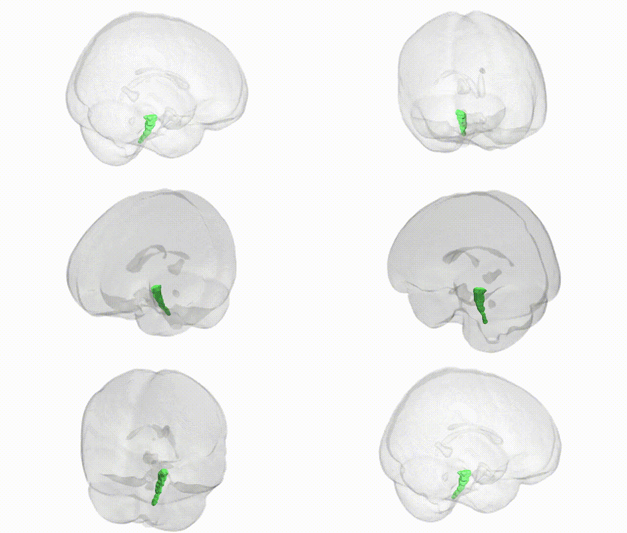
Full Quality Version: Download MP4
Wali Sidiqyar*, Gaurav Rudravaram*, Elyssa M. McMaster, Trent M. Schwartz, Adam M. Saunders, Kurt G. Schilling, Bennett A. Landman "Introducing SPINS: A Shared Public Visualization Library of Neuroanatomical Structures." Medical Imaging with Deep Learning- short paper
This resource is licensed under CC0 1.0 Universal (Public Domain).