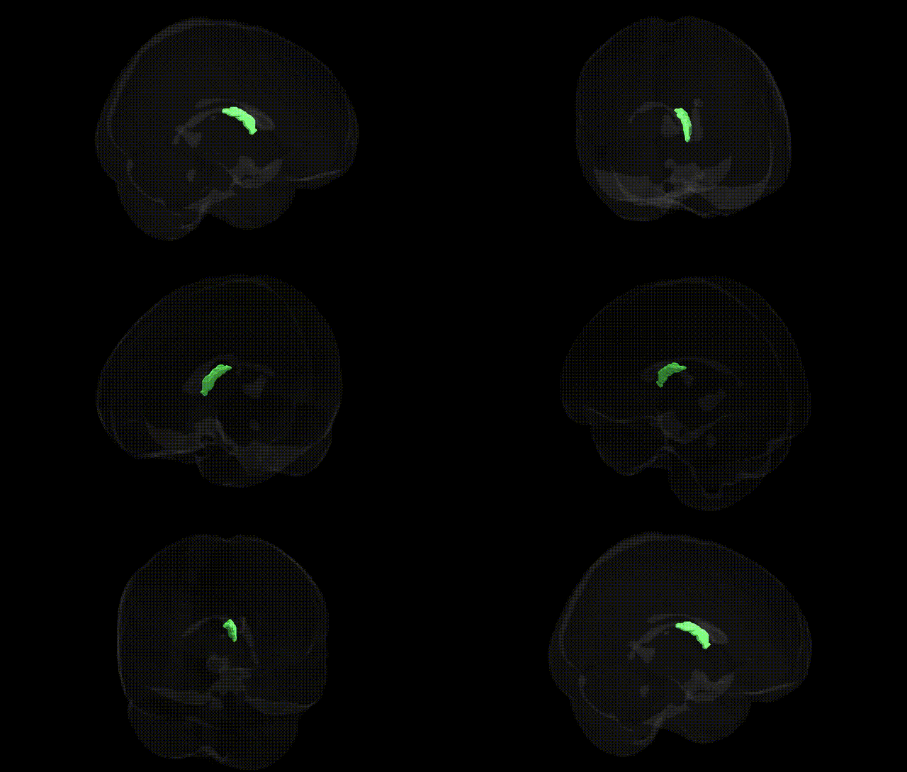
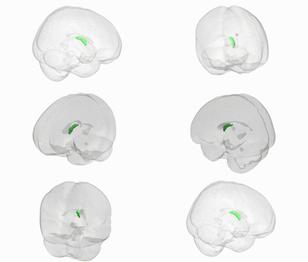
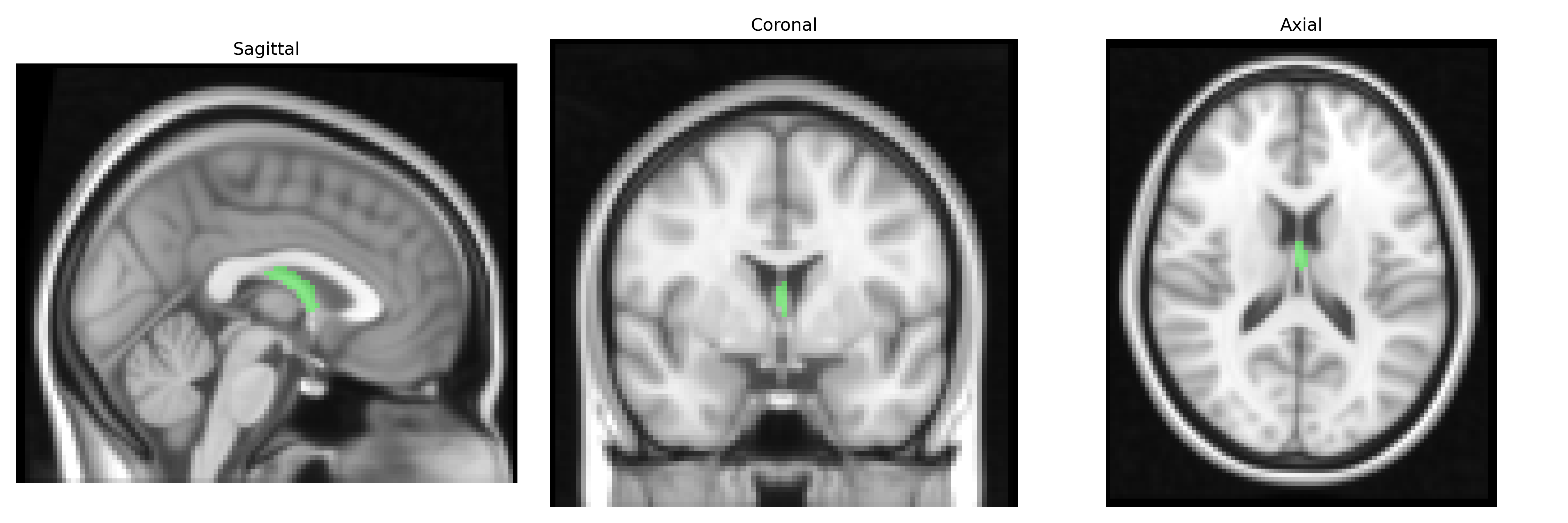
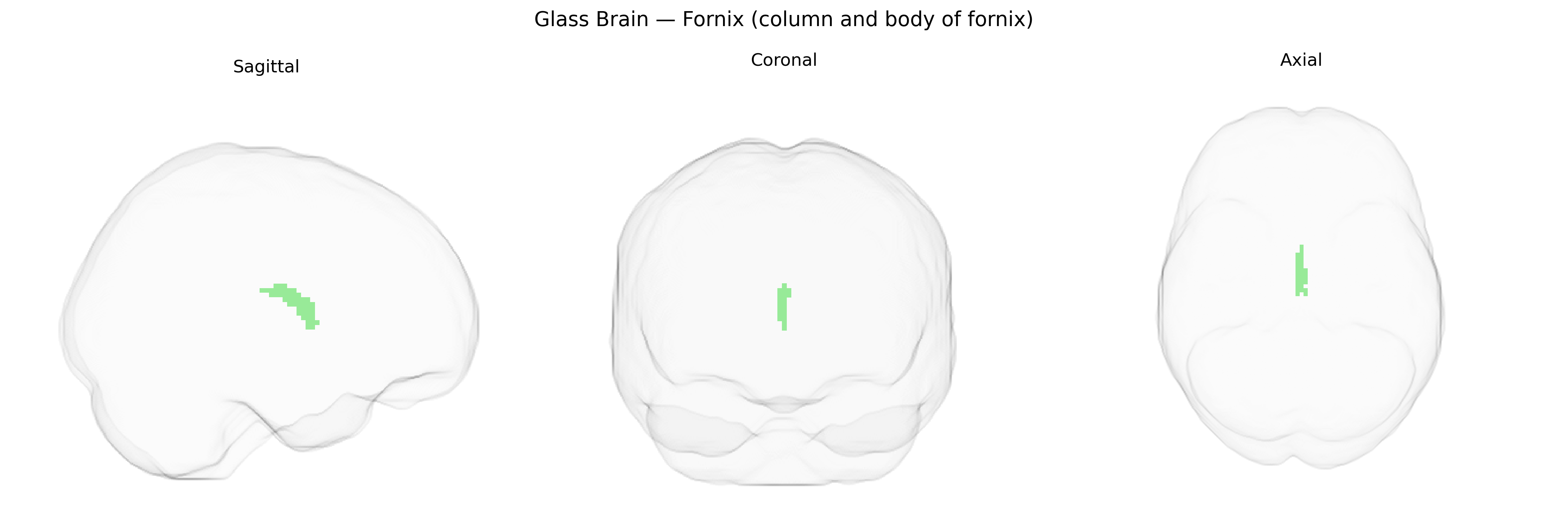
The bilateral fornix (column and body of fornix), as defined in the JHU ICBM 2mm Atlas, is a major white matter tract of the limbic system that arises primarily from the hippocampal formation and projects to the mammillary bodies, septal nuclei, and other hypothalamic and limbic structures. Anatomically, the body of the fornix consists of paired longitudinal fibers arching beneath the corpus callosum, while the columns descend anteriorly toward the diencephalon, forming a key conduit for hippocampal output. Functionally, this tract is critically involved in episodic memory processing, consolidation, and retrieval, and damage to the fornix is associated with profound anterograde amnesia and other memory impairments. As a central component of the Papez circuit and broader limbic networks, the fornix integrates hippocampal signals with subcortical regions to support mnemonic and affective functions. Fornix (brain)
The bilateral fornix (column and body) from the JHU ICBM labels 2 mm atlas, a core limbic white-matter tract connecting hippocampus and diencephalic structures, has been implicated in several genetic and GWAS-based associations, largely through diffusion MRI and imaging-genetics studies. Variants in genes affecting myelination and axonal integrity (e.g., NTRK1, MAG, PLP1, and broader oligodendrocyte-related pathways) have been reported to influence fornix fractional anisotropy (FA) and mean diffusivity, though specific loci often vary across cohorts. Fornix microstructure shows heritability in twin and family studies and has been associated with polygenic risk for Alzheimer’s disease and schizophrenia, with APOE ε4 status and other AD-risk variants linked to reduced fornix FA and volume, paralleling early limbic white-matter degeneration. GWAS of white-matter tracts (including UK Biobank–scale analyses) have identified loci that broadly affect limbic and hippocampal pathways—such as genes involved in axonal guidance, neurodevelopment, and extracellular matrix regulation—that partly map onto fornix metrics, although tract-specific hits for the fornix alone are less consistently replicated. Clinically, genetic liability for major depressive disorder, bipolar disorder, and schizophrenia has been associated with altered fornix integrity in imaging-genetics studies, and rare monogenic conditions impacting midline commissural and limbic development (e.g., certain callosal and septal agenesis syndromes) can produce marked fornix abnormalities, underscoring the tract’s sensitivity to neurodevelopmental genetic variation.
Overview generated by GPT-4o (2026).
Region ID: 6
Hemisphere: bilateral
Atlas: JHU ICBM labels 2mm
Full Quality Version: Download MP4
Full Quality Version: Download MP4
Wali Sidiqyar*, Gaurav Rudravaram*, Elyssa M. McMaster, Trent M. Schwartz, Adam M. Saunders, Kurt G. Schilling, Bennett A. Landman "Introducing SPINS: A Shared Public Visualization Library of Neuroanatomical Structures." Medical Imaging with Deep Learning- short paper
This resource is licensed under CC0 1.0 Universal (Public Domain).