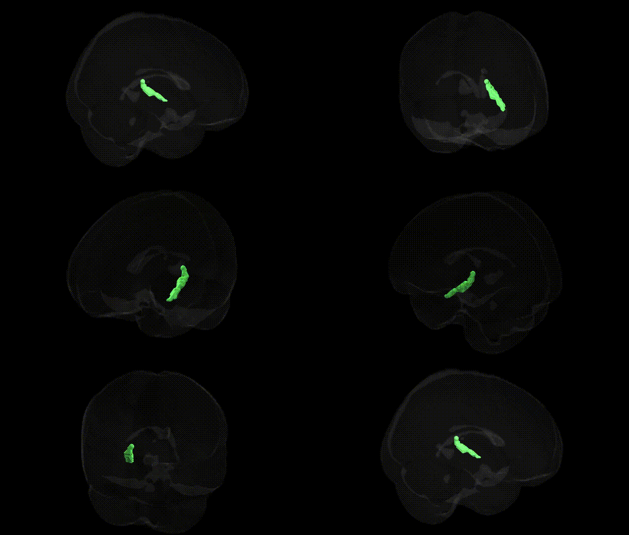
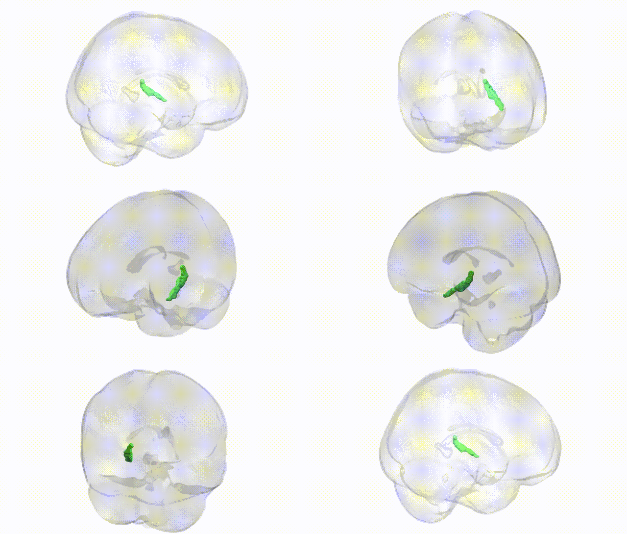
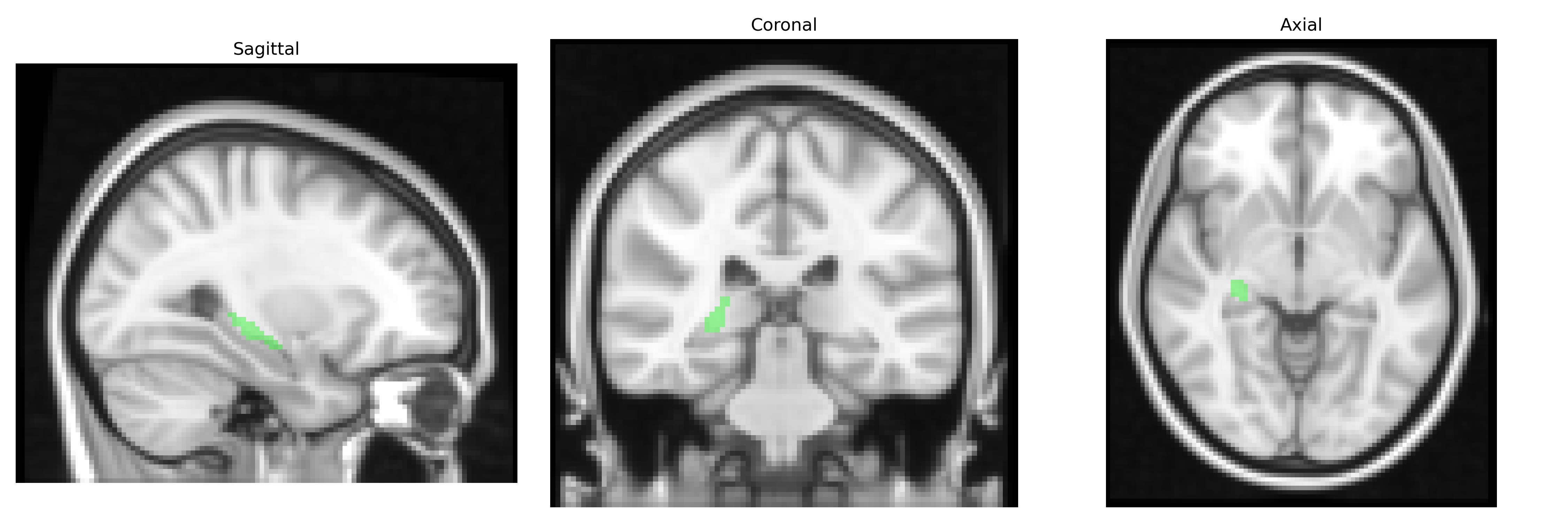
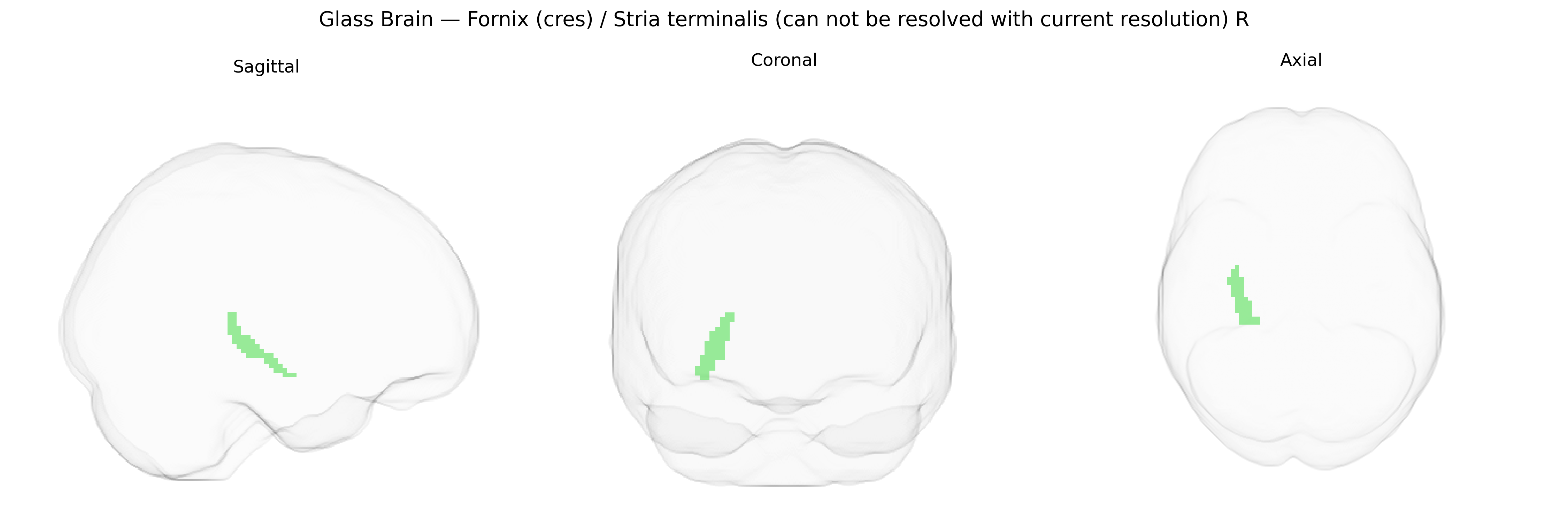
The bilateral fornix (cres)/stria terminalis (cannot be resolved with current resolution) R, as defined in the JHU ICBM 2 mm atlas, represents a composite label encompassing two closely apposed limbic fiber bundles that cannot be reliably distinguished at this spatial resolution. The fornix is a major efferent white matter pathway of the hippocampal formation, arching beneath the corpus callosum and projecting primarily to the mammillary bodies and septal nuclei, thereby supporting episodic memory and hippocampal–diencephalic communication. The stria terminalis courses along the margin of the thalamus, linking the amygdala with the hypothalamus and septal regions, and is a key conduit for emotional, autonomic, and neuroendocrine regulation. In diffusion MRI and structural atlases, these tracts overlap in periventricular and medial temporal regions, leading to their joint labeling as a single region of interest in probabilistic white matter atlases such as the JHU ICBM framework. Fornix (neuroanatomy)
Genetic associations involving the right fornix/stria terminalis region, as defined in the JHU ICBM 2 mm atlas, largely emerge from imaging-genetics and GWAS of white-matter microstructure and limbic circuitry rather than from region-specific candidate-gene work. Diffusion MRI GWAS have identified common variants near genes implicated in axon guidance, myelination, and neurodevelopment (such as NTRK1/2, MAG, and various cell-adhesion and cytoskeletal genes) that influence fractional anisotropy and other diffusion metrics in the fornix and adjacent limbic tracts, with some loci overlapping those implicated in hippocampal volume and broader limbic network structure. Polygenic risk for Alzheimer’s disease, schizophrenia, and major depression has been associated with altered fornix integrity, consistent with the tract’s role in memory and emotional regulation; APOE ε4 and other AD risk variants show convergent associations with reduced fornix microstructural integrity in older adults and prodromal AD, while schizophrenia and psychosis risk variants (including in genes related to glutamatergic signaling and synaptic regulation) have been linked to abnormalities in fornix and stria terminalis pathways in case–control and PRS-based imaging studies. Additional genetic links arise from GWAS of anxiety, PTSD, and stress-related traits, where risk alleles in HPA-axis and neurotrophin-related genes show indirect associations with structural or functional alterations in amygdala–fornix/stria terminalis circuits, though these findings are generally at the network or tract level and not specific to the right hemisphere label in the JHU atlas.
Overview generated by GPT-4o (2026).
Region ID: 39
Hemisphere: bilateral
Atlas: JHU ICBM labels 2mm
Full Quality Version: Download MP4
Full Quality Version: Download MP4
Wali Sidiqyar*, Gaurav Rudravaram*, Elyssa M. McMaster, Trent M. Schwartz, Adam M. Saunders, Kurt G. Schilling, Bennett A. Landman "Introducing SPINS: A Shared Public Visualization Library of Neuroanatomical Structures." Medical Imaging with Deep Learning- short paper
This resource is licensed under CC0 1.0 Universal (Public Domain).