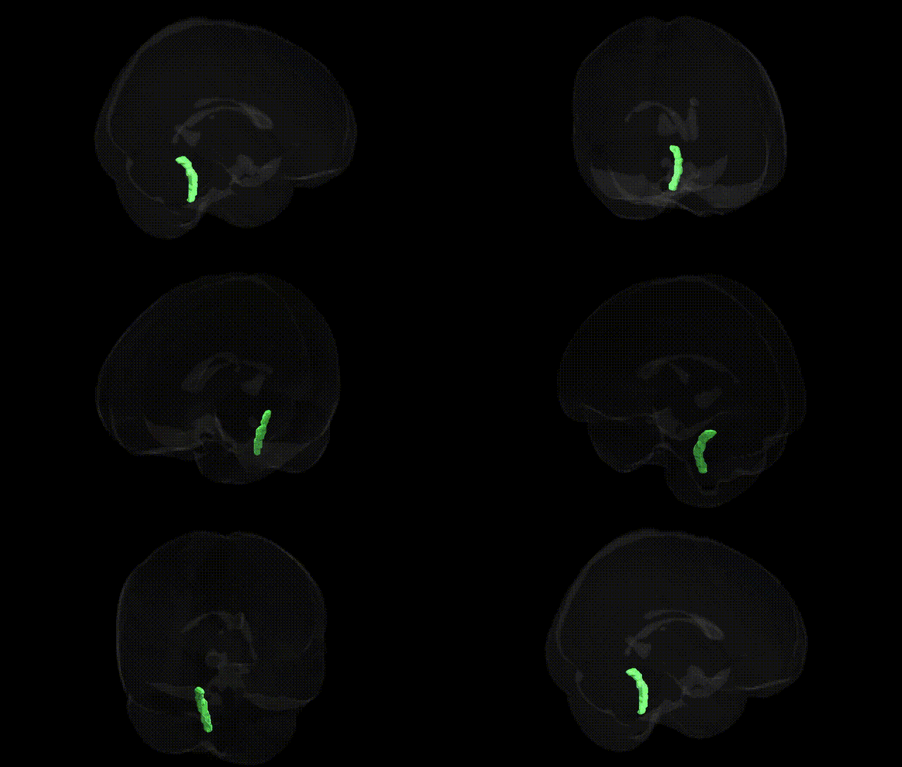
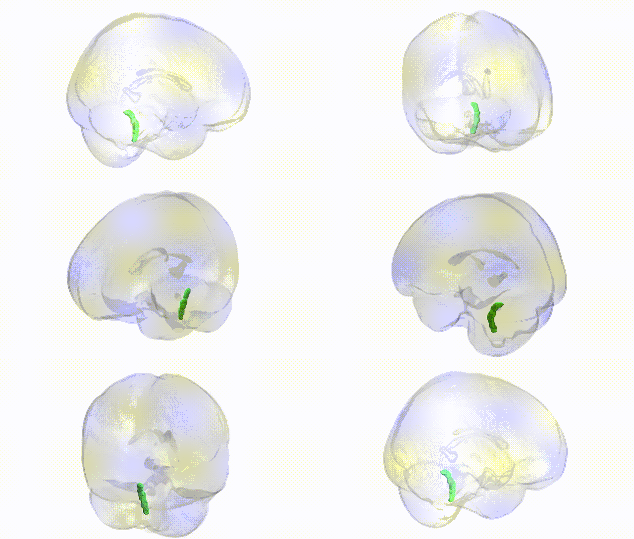
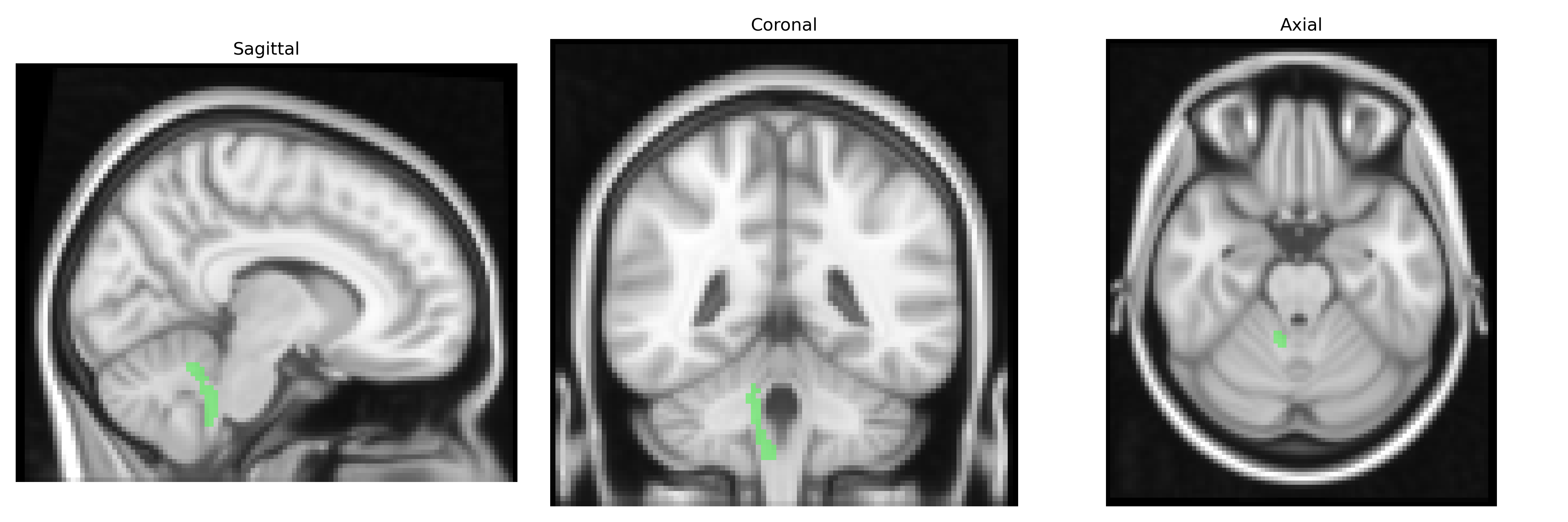
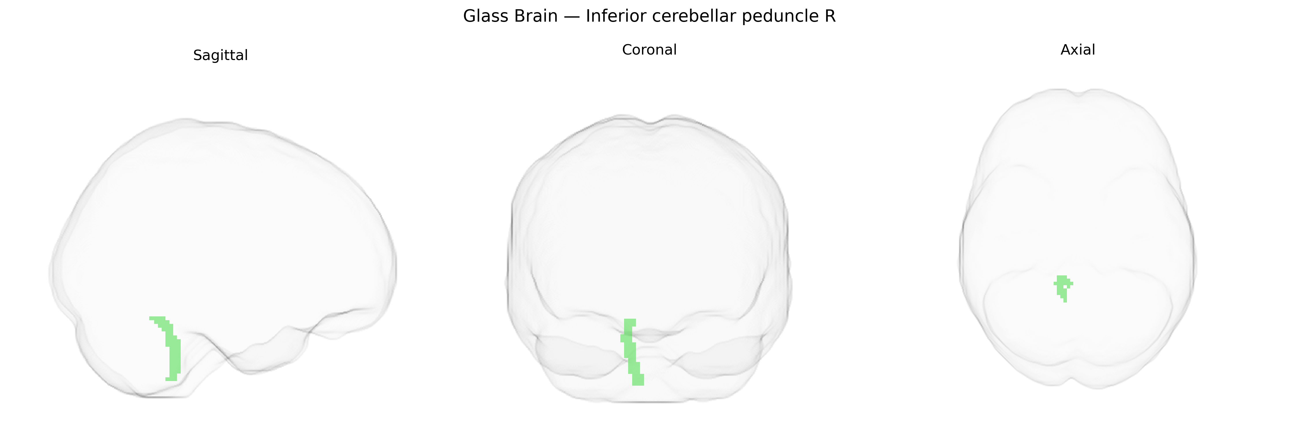
The bilateral Inferior cerebellar peduncle (right) is a major white matter tract connecting the medulla oblongata to the cerebellum, carrying predominantly afferent fibers that convey proprioceptive and vestibular information essential for coordination, balance, and motor learning. It includes inputs from the dorsal spinocerebellar tract, cuneocerebellar fibers, olivocerebellar fibers from the inferior olivary nucleus, and vestibulocerebellar projections, integrating peripheral sensory signals with cerebellar circuits that fine-tune posture and movement. This structure plays a critical role in maintaining equilibrium and adjusting motor activity in response to changing sensory feedback, and lesions in this pathway can result in ataxia, dysmetria, and other cerebellar signs. There is no direct link for the inferior cerebellar peduncle; a closely related structure is the Cerebellum.
The bilateral inferior cerebellar peduncle (right) as defined in the JHU ICBM 2 mm atlas is part of the major cerebellar afferent pathway, and genetic associations involving this tract typically come from imaging genetics and diffusion MRI GWAS rather than region-specific candidate gene studies. Large-scale brain-wide GWAS of white matter microstructure have identified polygenic influences on fractional anisotropy and other diffusion metrics in cerebellar peduncles, implicating common variants in genes involved in axon guidance, myelination, and neurodevelopmental processes (for example, loci near genes such as CNTN4, ROBO2, and PLP1 in broader cerebellar and brainstem tracts), though findings are usually reported at the level of cerebellar peduncles or brainstem tracts collectively rather than the right inferior cerebellar peduncle alone. Imaging genetics studies in disorders such as multiple sclerosis, hereditary ataxias, and neurodevelopmental conditions (including autism spectrum disorder and ADHD) have associated risk variants with altered cerebellar white matter, including inferior cerebellar peduncle involvement, but these links are typically indirect, reflecting global cerebellar or brainstem changes rather than a tract-specific GWAS signal. Overall, current genetic evidence supports a polygenic architecture influencing microstructural variation in cerebellar peduncles and their susceptibility in neurodegenerative and neurodevelopmental disorders, but there are no well-established, unique genetic loci that have been robustly and specifically tied to the bilateral inferior cerebellar peduncle (R) region in the JHU ICBM atlas.
Overview generated by GPT-4o (2026).
Region ID: 11
Hemisphere: bilateral
Atlas: JHU ICBM labels 2mm
Full Quality Version: Download MP4
Full Quality Version: Download MP4
Wali Sidiqyar*, Gaurav Rudravaram*, Elyssa M. McMaster, Trent M. Schwartz, Adam M. Saunders, Kurt G. Schilling, Bennett A. Landman "Introducing SPINS: A Shared Public Visualization Library of Neuroanatomical Structures." Medical Imaging with Deep Learning- short paper
This resource is licensed under CC0 1.0 Universal (Public Domain).