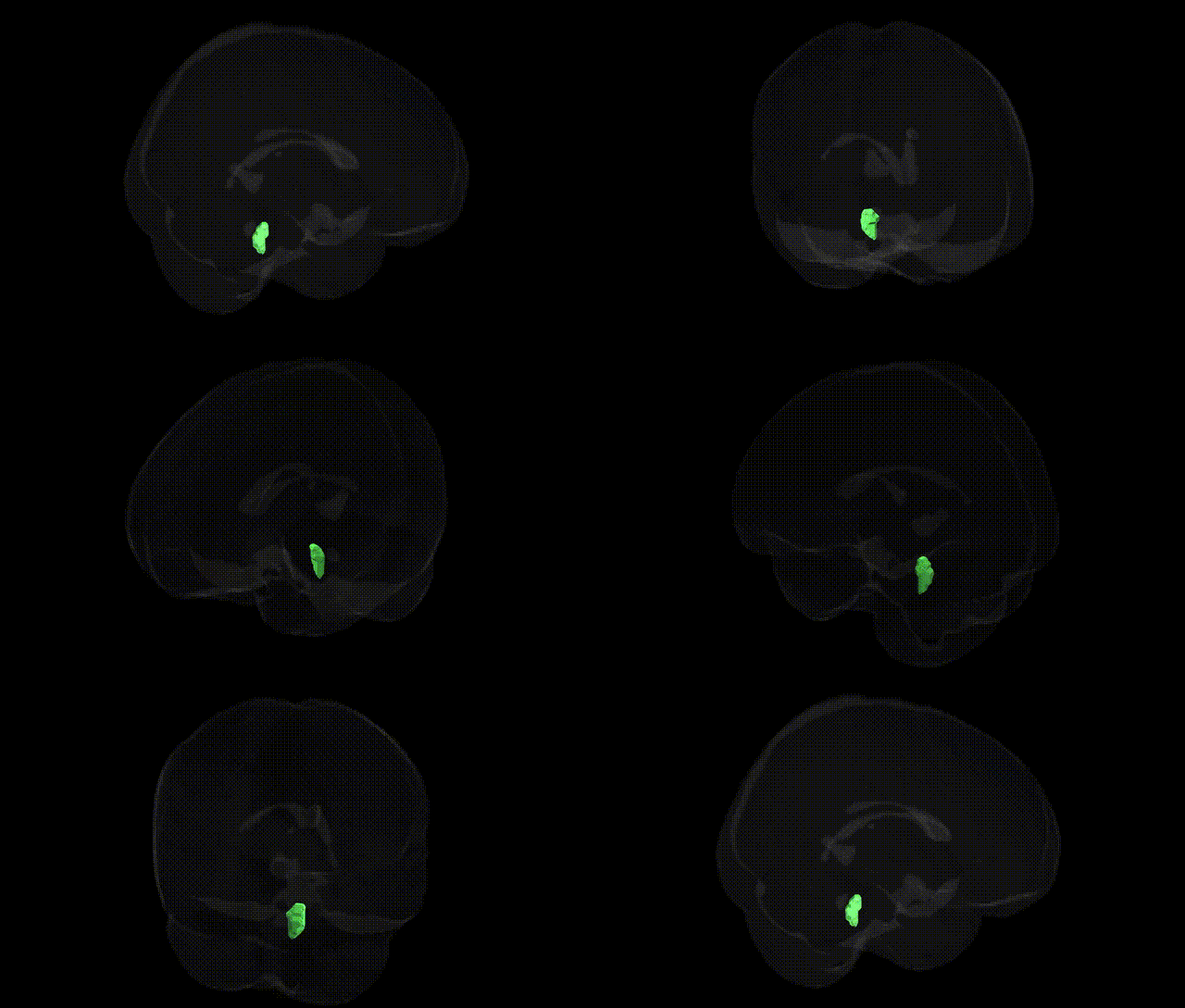
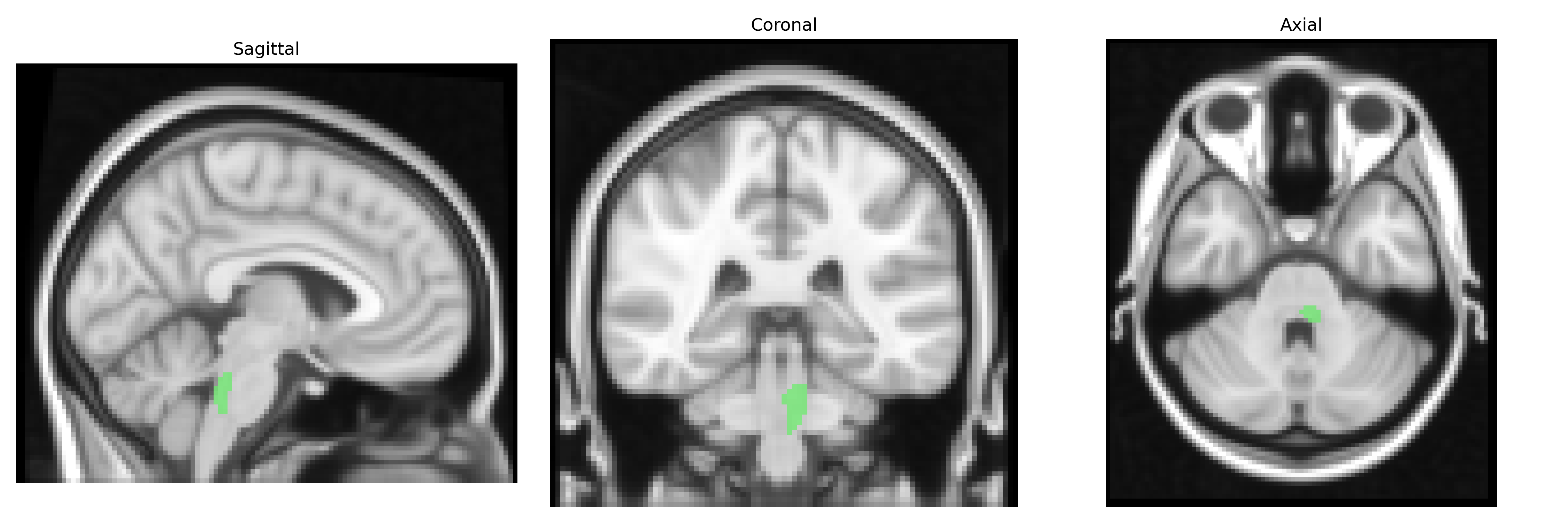
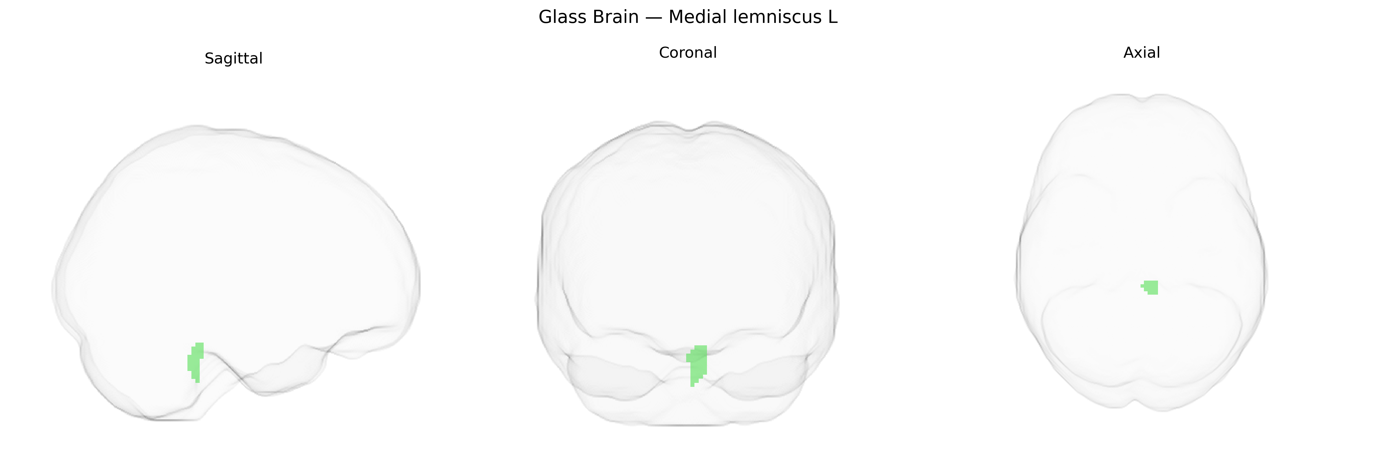
The bilateral Medial lemniscus L, as defined in the JHU ICBM labels 2mm atlas, corresponds to the left-sided component of the medial lemniscus, a major ascending somatosensory pathway in the brainstem. It carries highly processed tactile and proprioceptive information from the contralateral body, originating from second-order neurons in the dorsal column nuclei (gracile and cuneate nuclei) of the medulla and projecting rostrally to the ventral posterolateral nucleus of the thalamus. This tract is somatotopically organized and composed of heavily myelinated axons, supporting rapid transmission of fine touch, vibration, and position sense. Functionally, integrity of the medial lemniscus is essential for discriminative somatosensation and contributes to conscious perception of body position and detailed tactile features. There is no direct Wikipedia article for this exact atlas label; a closely related structure is described here: Medial lemniscus.
The bilateral medial lemniscus (L) region from the JHU ICBM labels 2mm atlas is a brainstem white matter tract that primarily carries somatosensory information and, as such, has not been a common focus of region-specific genetic association studies. Large-scale GWAS and imaging-genetics work (e.g., ENIGMA consortium, UK Biobank) have identified numerous loci associated with global and regional white matter microstructure, often measured via diffusion tensor imaging, but these findings typically map to broader tracts (such as major commissural and association pathways) rather than specifically isolating the medial lemniscus. Some variants in genes related to axon guidance, myelination, and neurodevelopment (for example, in pathways involving cell adhesion molecules and oligodendrocyte function) have been associated with white matter integrity measures and neurological or psychiatric conditions, yet these studies rarely report discrete, robust associations localized to the medial lemniscus itself. Disorders that affect somatosensory pathways—such as hereditary spastic paraplegias, spinocerebellar ataxias, and certain leukodystrophies—can involve degeneration or abnormal development of brainstem tracts, including those encompassing the medial lemniscus, but the genetic findings pertain to the disease as a whole rather than to this specific atlas-defined region, and current GWAS literature does not provide well-established, trait-specific genetic associations uniquely attributed to the bilateral medial lemniscus (L) in the JHU ICBM framework.
Overview generated by GPT-4o (2026).
Region ID: 10
Hemisphere: bilateral
Atlas: JHU ICBM labels 2mm
Full Quality Version: Download MP4
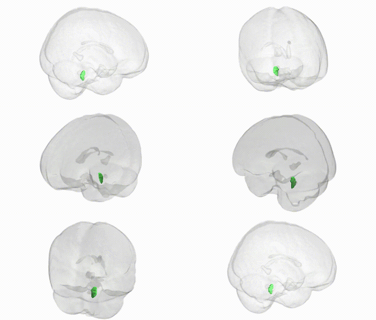
Full Quality Version: Download MP4
Wali Sidiqyar*, Gaurav Rudravaram*, Elyssa M. McMaster, Trent M. Schwartz, Adam M. Saunders, Kurt G. Schilling, Bennett A. Landman "Introducing SPINS: A Shared Public Visualization Library of Neuroanatomical Structures." Medical Imaging with Deep Learning- short paper
This resource is licensed under CC0 1.0 Universal (Public Domain).