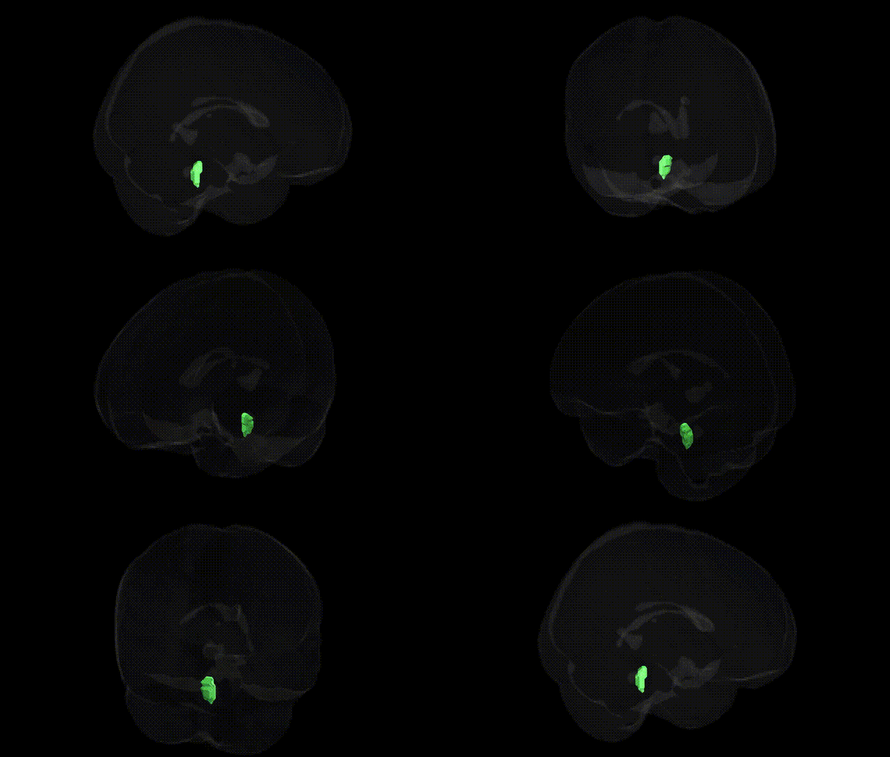
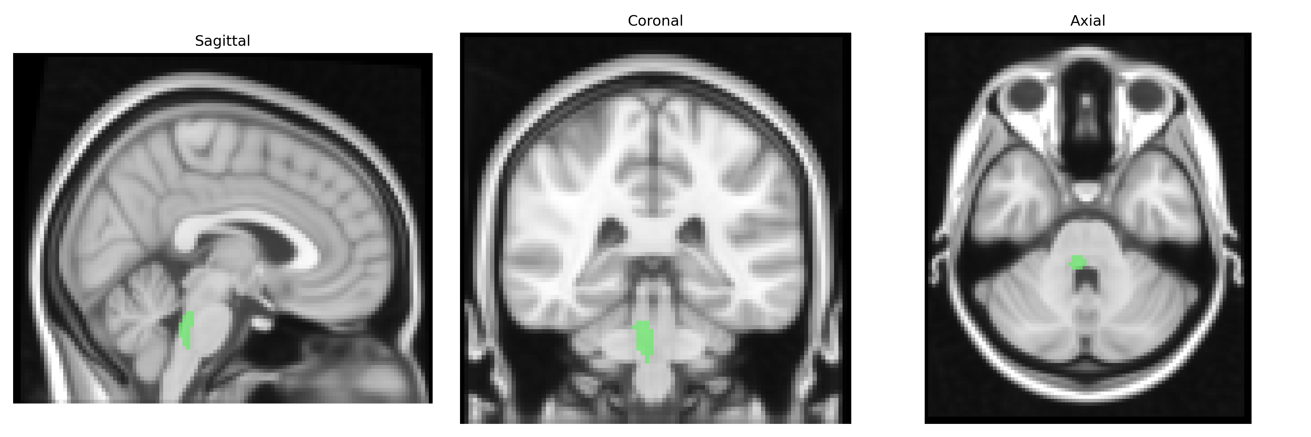
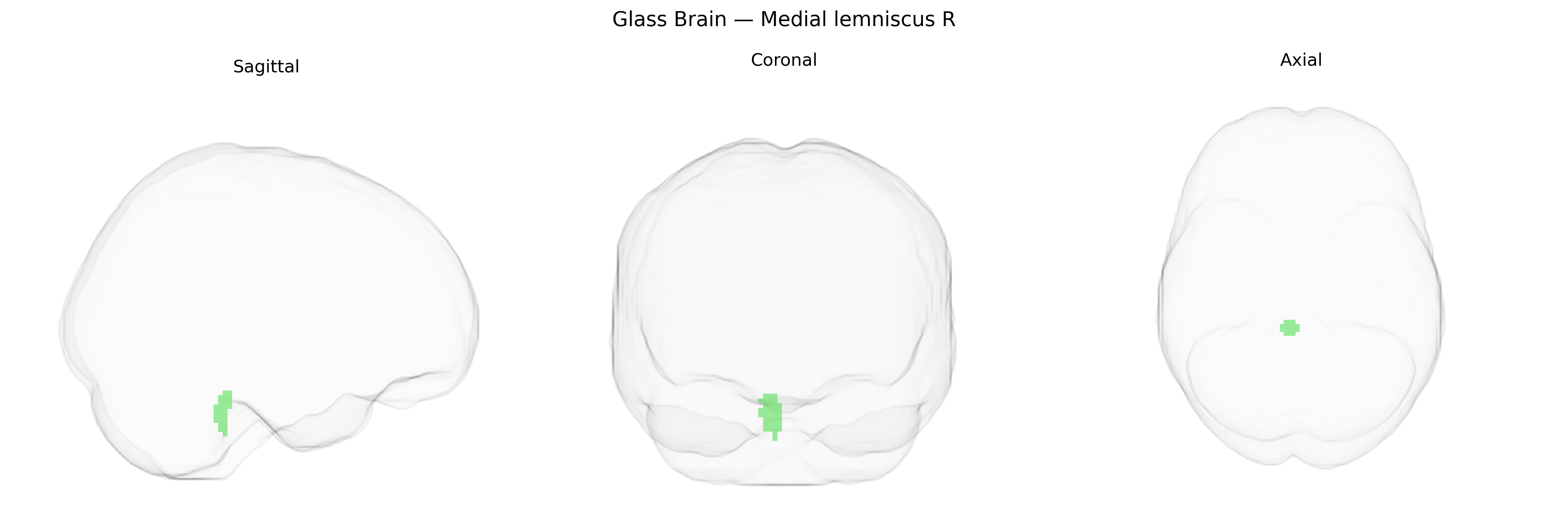
The bilateral Medial lemniscus R, as defined in the JHU ICBM 2 mm atlas, corresponds to the right-sided component of the medial lemniscus, a major ascending somatosensory pathway in the brainstem that conveys information about fine touch, vibration, and proprioception from the contralateral body to higher cortical centers. Originating from second-order neurons in the dorsal column nuclei (gracile and cuneate nuclei) of the medulla, axons decussate and ascend within the medial lemniscus through the medulla, pons, and midbrain, maintaining a somatotopic organization before synapsing in the ventral posterolateral nucleus of the thalamus. From there, third-order neurons project to the primary somatosensory cortex, allowing conscious perception of discriminative touch and limb position. The medial lemniscus is thus integral to the dorsal column–medial lemniscus system and is functionally critical for precise tactile and proprioceptive processing. Medial lemniscus
The bilateral medial lemniscus, as defined in the JHU ICBM 2 mm atlas, is a brainstem white-matter tract that transmits somatosensory information from the dorsal column nuclei to the thalamus, and direct, region-specific genetic associations from GWAS are currently sparse, reflecting the underrepresentation of brainstem pathways in large neuroimaging-genetics consortia. Diffusion MRI–based studies combining GWAS with tract-specific measures (e.g., fractional anisotropy and mean diffusivity) have more frequently implicated major supratentorial tracts, but some polygenic influences on general white-matter microstructure and brainstem integrity—such as variants in genes involved in axon guidance (e.g., ROBO, SLIT family), myelination (e.g., MAG, MOBP, PLP1), and neurodevelopmental signaling pathways—are likely to affect the medial lemniscus indirectly as part of global or brainstem-wide effects. Clinical genetics literature more often links the function of medial lemniscus pathways to disease via broader brainstem or somatosensory circuitry: Mendelian and risk variants for neurodegenerative and demyelinating disorders (including Parkinson’s disease, multiple system atrophy, and multiple sclerosis) have been associated with brainstem and posterior column degeneration that would encompass the medial lemniscus, and sensory abnormalities in hereditary neuropathies and spinocerebellar ataxias may involve its upstream and downstream relays. Large psychiatric and cognitive GWAS (e.g., for schizophrenia, depression, intelligence, and educational attainment) sometimes report associations with brainstem or global white-matter measures but do not specifically isolate the medial lemniscus, making any genetic inferences about this tract extrapolations from broader somatosensory and brainstem networks rather than direct, tract-level findings.
Overview generated by GPT-4o (2026).
Region ID: 9
Hemisphere: bilateral
Atlas: JHU ICBM labels 2mm
Full Quality Version: Download MP4
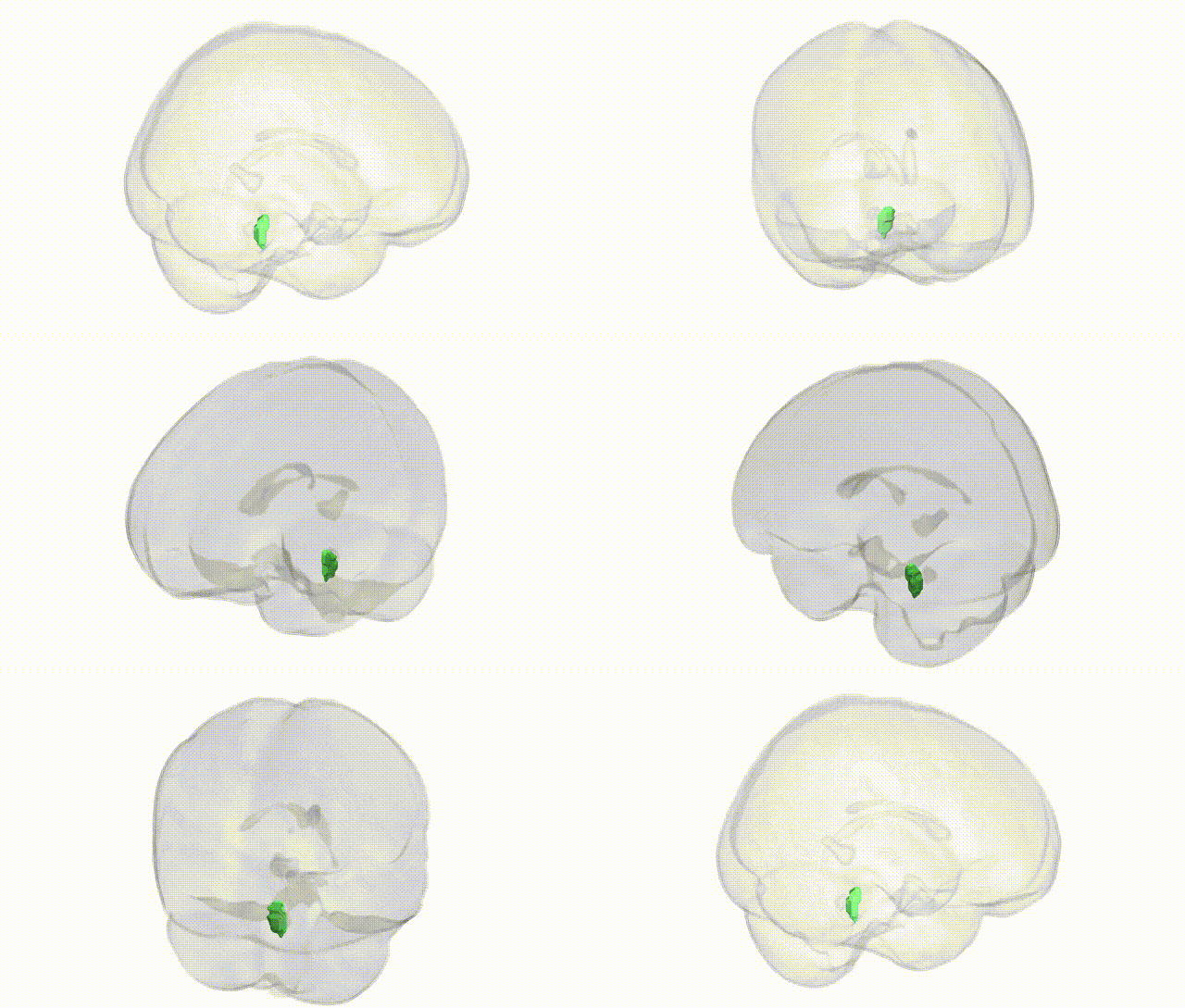
Full Quality Version: Download MP4
Wali Sidiqyar*, Gaurav Rudravaram*, Elyssa M. McMaster, Trent M. Schwartz, Adam M. Saunders, Kurt G. Schilling, Bennett A. Landman "Introducing SPINS: A Shared Public Visualization Library of Neuroanatomical Structures." Medical Imaging with Deep Learning- short paper
This resource is licensed under CC0 1.0 Universal (Public Domain).