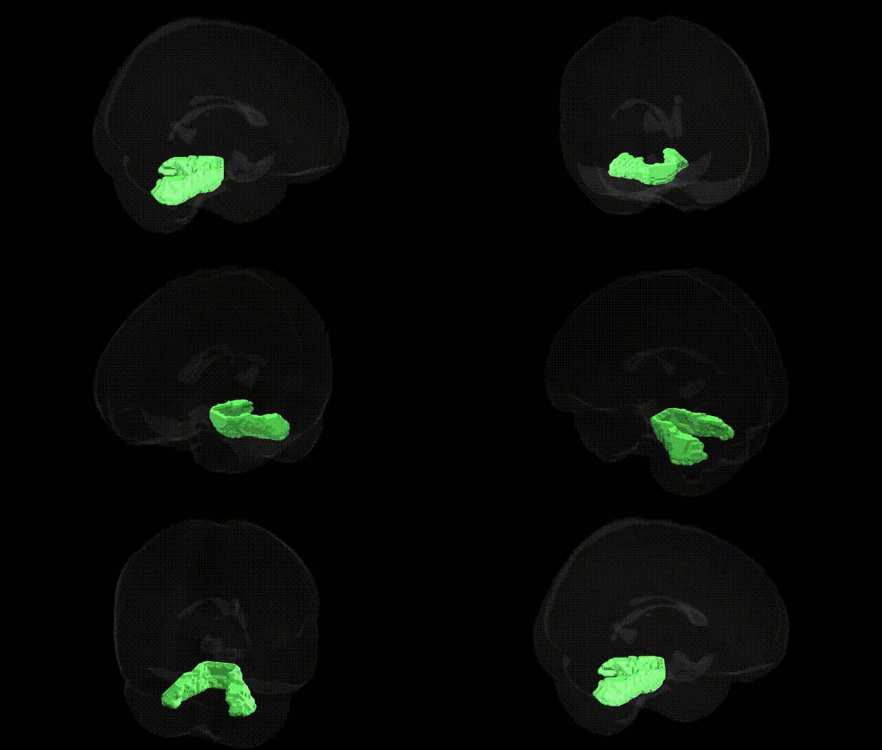
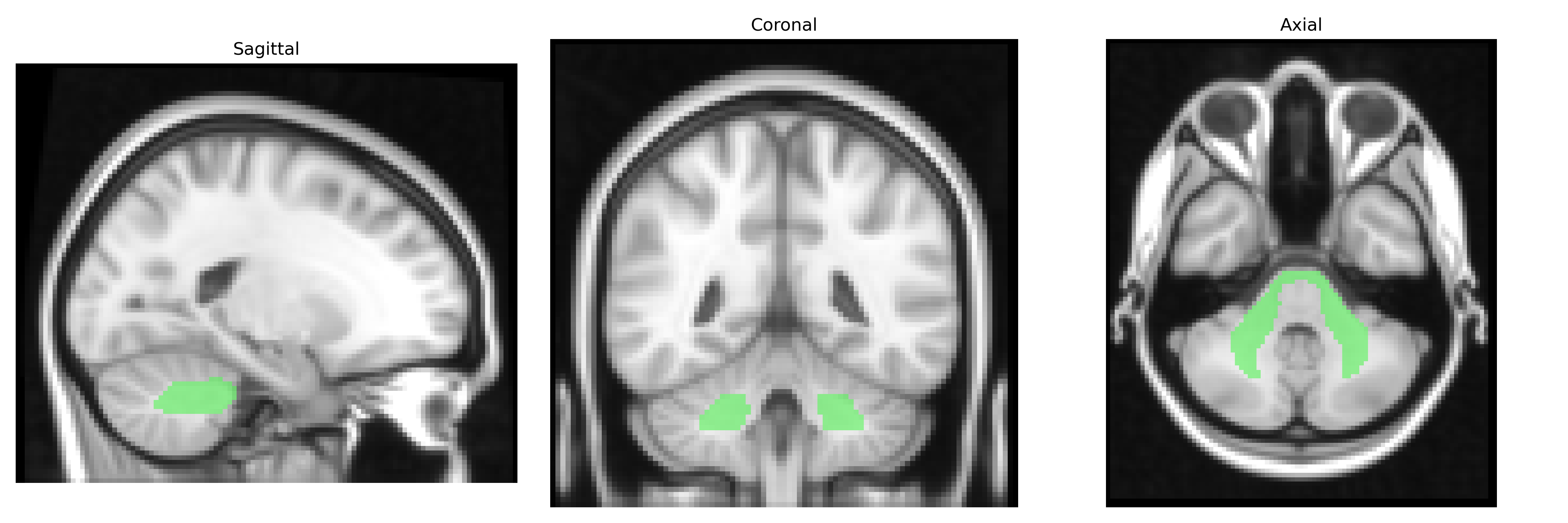
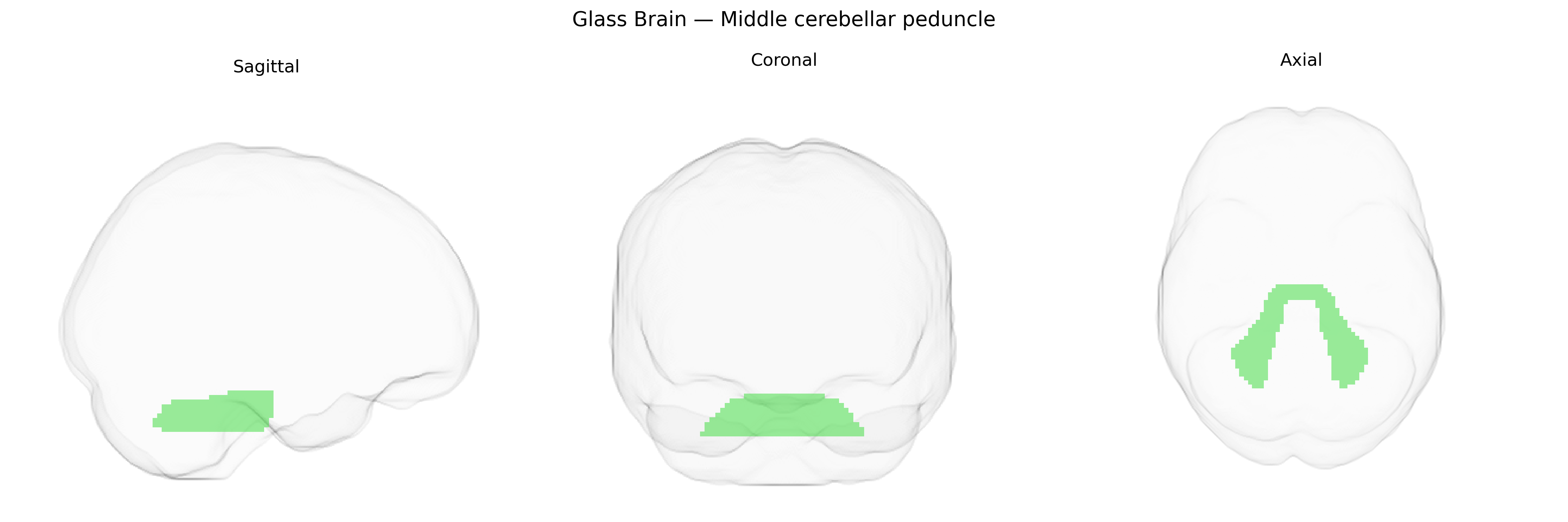
The bilateral Middle cerebellar peduncle is a paired white-matter structure that forms the largest of the cerebellar peduncles, consisting primarily of massive transverse pontocerebellar fibers that convey information from the contralateral cerebral cortex (via pontine nuclei) to the cerebellar hemispheres. Located laterally on each side of the pons, it serves as a major afferent pathway to the cerebellum, playing a central role in the coordination of voluntary movement, motor learning, and the integration of cortical motor plans with proprioceptive feedback. Damage or degeneration of the middle cerebellar peduncle is associated with cerebellar ataxia, dysmetria, and gait disturbance, and can be seen in conditions such as multiple system atrophy and certain leukodystrophies. Middle cerebellar peduncle
The bilateral middle cerebellar peduncle (MCP), as defined in the JHU ICBM-DTI-81 2mm atlas, has been implicated in several genetic and neuropsychiatric contexts, although direct, region-specific genetic associations remain limited and largely derived from imaging-genetics and DTI-based GWAS of white matter microstructure. Polygenic influences on MCP fractional anisotropy and related diffusion metrics have been reported in large cohorts such as UK Biobank and ENIGMA-related analyses, indicating substantial heritability and shared genetic architecture with general brain white matter integrity. MCP integrity has been associated with genetic risk for schizophrenia, bipolar disorder, and major depressive disorder in imaging-genetics studies, where higher polygenic risk scores for these disorders correlate with altered diffusion measures in cerebellar and brainstem tracts including the MCP. Variants in genes involved in axonal guidance, myelination, and oligodendrocyte function (e.g., NTRK1/2-related pathways, CNTN4/6, and myelin-related loci identified in white matter GWAS) have been indirectly linked to MCP structure as part of global white matter effects rather than MCP-specific hits. In neurodevelopmental and neurodegenerative conditions, MCP abnormalities have been noted in individuals with copy-number variants such as 22q11.2 deletion syndrome and in spinocerebellar ataxias and multiple system atrophy, where pathogenic variants in ataxia-related genes (e.g., ATXN1, ATXN2, ATXN3, CACNA1A) and synuclein/oligodendroglial pathways contribute to pronounced MCP atrophy or signal change, but these are typically described at the syndrome level rather than as focal MCP GWAS associations. Overall, evidence supports that the MCP is a heritable white matter structure affected by polygenic influences on brain connectivity and by monogenic or CNV-driven disorders that disrupt cerebello-cerebral circuits, though robust, MCP-specific genetic loci from unbiased GWAS remain sparse.
Overview generated by GPT-4o (2026).
Region ID: 1
Hemisphere: bilateral
Atlas: JHU ICBM labels 2mm
Full Quality Version: Download MP4
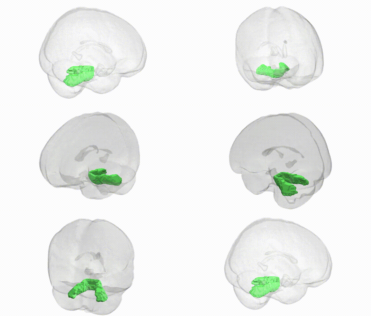
Full Quality Version: Download MP4
Wali Sidiqyar*, Gaurav Rudravaram*, Elyssa M. McMaster, Trent M. Schwartz, Adam M. Saunders, Kurt G. Schilling, Bennett A. Landman "Introducing SPINS: A Shared Public Visualization Library of Neuroanatomical Structures." Medical Imaging with Deep Learning- short paper
This resource is licensed under CC0 1.0 Universal (Public Domain).