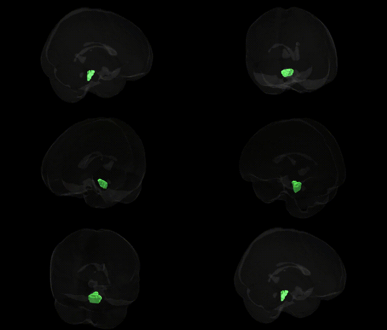
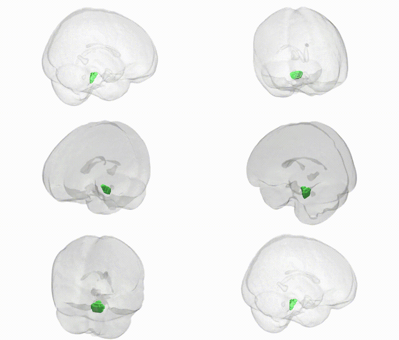
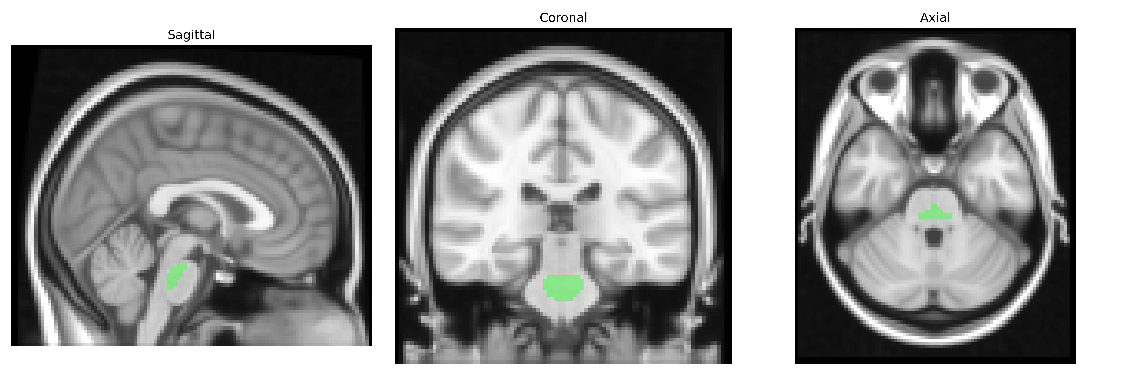
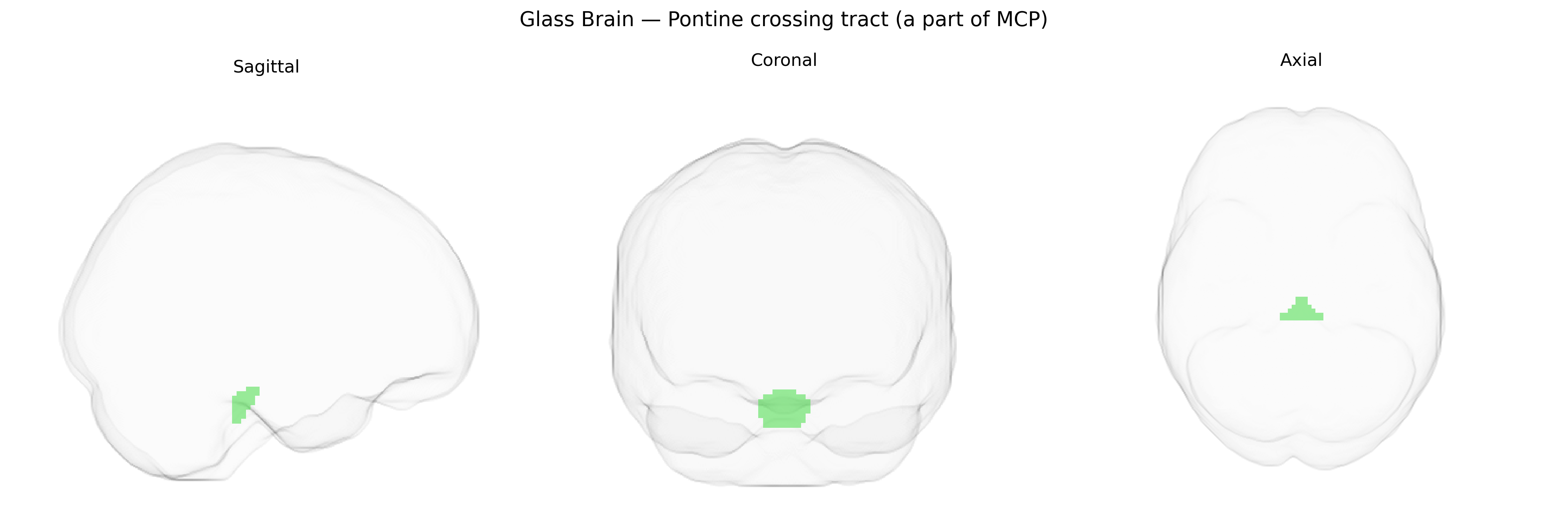
The bilateral Pontine crossing tract, a major component of the middle cerebellar peduncle, consists predominantly of transverse pontine fibers arising from pontine nuclei that decussate in the pons and project to the contralateral cerebellar hemisphere. These fibers form a critical conduit within the cortico-ponto-cerebellar pathway, transmitting information derived from widespread cerebral cortical areas to the cerebellar cortex, thereby supporting motor coordination, timing, and aspects of motor learning and sensorimotor integration. Anatomically, the pontine crossing tract occupies the ventral (basilar) portion of the pons and continues laterally into the middle cerebellar peduncle, contributing substantially to its bulk and characteristic transverse fiber architecture. There is no direct Wikipedia article for the “Pontine crossing tract”; a closely related structure is the Middle cerebellar peduncle.
The bilateral pontine crossing tract, a component of the middle cerebellar peduncle in the JHU ICBM 2mm atlas, has been implicated in several imaging-genetics and GWAS-based studies, although associations are often reported under broader labels such as “middle cerebellar peduncle” or “brainstem/cerebellar white matter.” Large-scale diffusion MRI GWAS (e.g., UK Biobank) have identified common variants in genes involved in axon guidance, myelination, and neuronal development (including loci near NCAN, LINGO1, and genes in the contactin/NRG–ErbB pathways) that influence microstructural properties like fractional anisotropy in this tract. Polygenic risk for schizophrenia, major depressive disorder, and bipolar disorder shows associations with altered integrity of pontocerebellar pathways, including the middle cerebellar peduncle, and rare variant or copy-number studies in neurodevelopmental disorders (autism spectrum disorder, intellectual disability, and certain ataxia syndromes) often report abnormalities in pontine–cerebellar white matter, though typically without tract-specific genetic resolution. Imaging-genetic work on motor coordination, cognitive performance, and gait-related traits has linked variants in genes affecting cerebellar and brainstem development (for example, ROBO/SLIT guidance genes and some cerebellar ataxia loci) to structural differences in this region, but current evidence remains indirect and usually embedded within broader brainstem or cerebellar phenotypes rather than uniquely pinpointing the bilateral pontine crossing tract.
Overview generated by GPT-4o (2026).
Region ID: 2
Hemisphere: bilateral
Atlas: JHU ICBM labels 2mm
Full Quality Version: Download MP4
Full Quality Version: Download MP4
Wali Sidiqyar*, Gaurav Rudravaram*, Elyssa M. McMaster, Trent M. Schwartz, Adam M. Saunders, Kurt G. Schilling, Bennett A. Landman "Introducing SPINS: A Shared Public Visualization Library of Neuroanatomical Structures." Medical Imaging with Deep Learning- short paper
This resource is licensed under CC0 1.0 Universal (Public Domain).