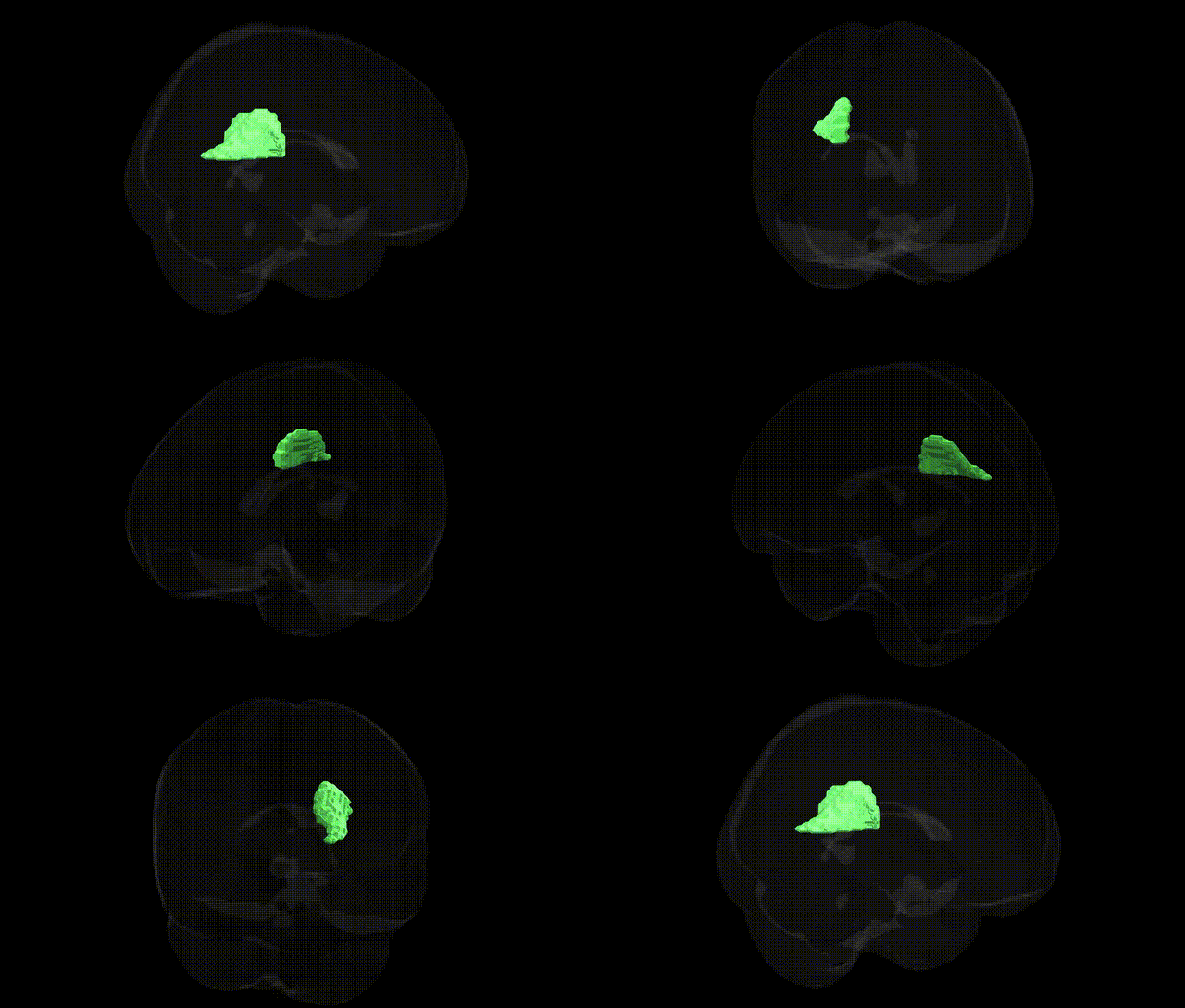
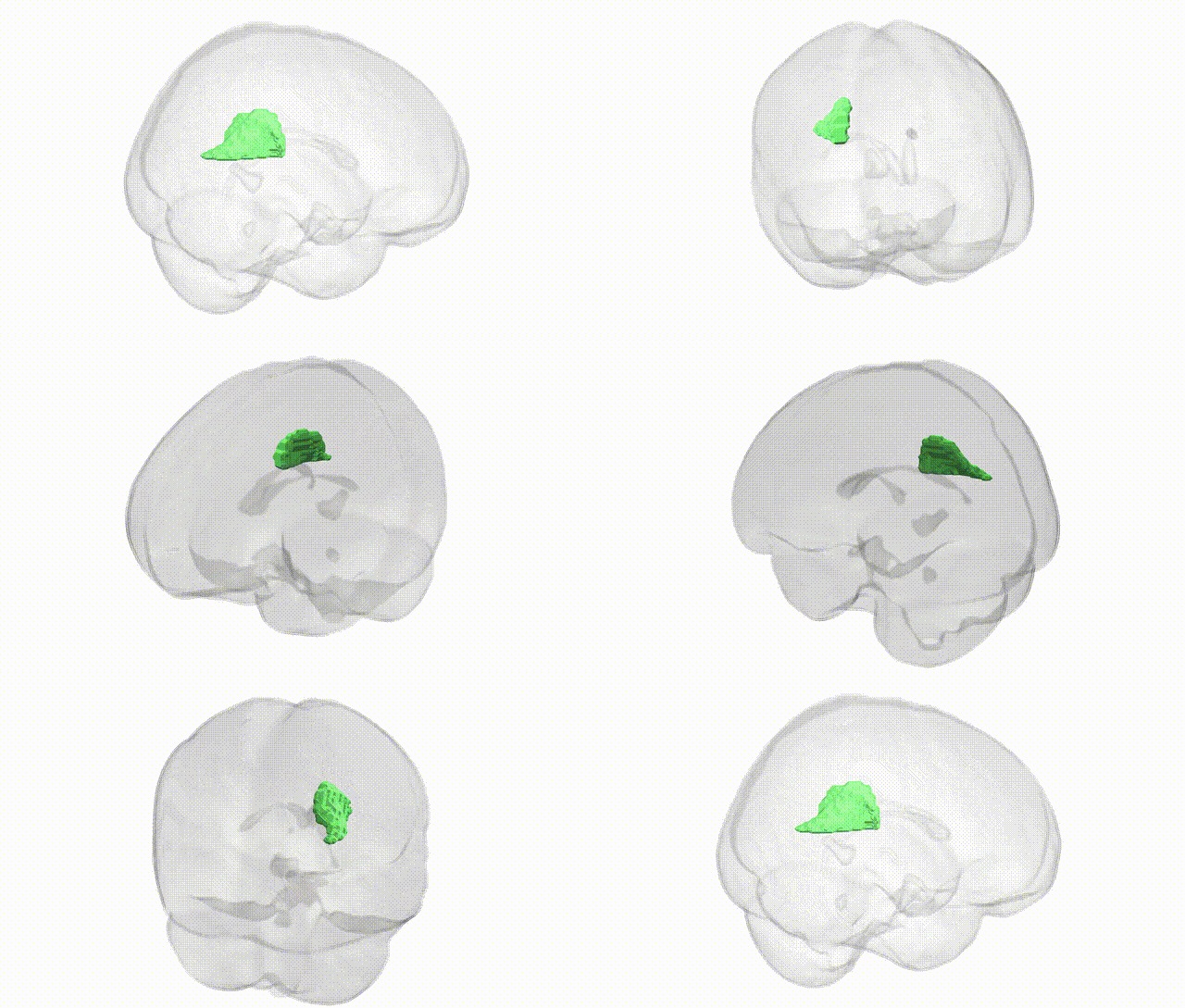
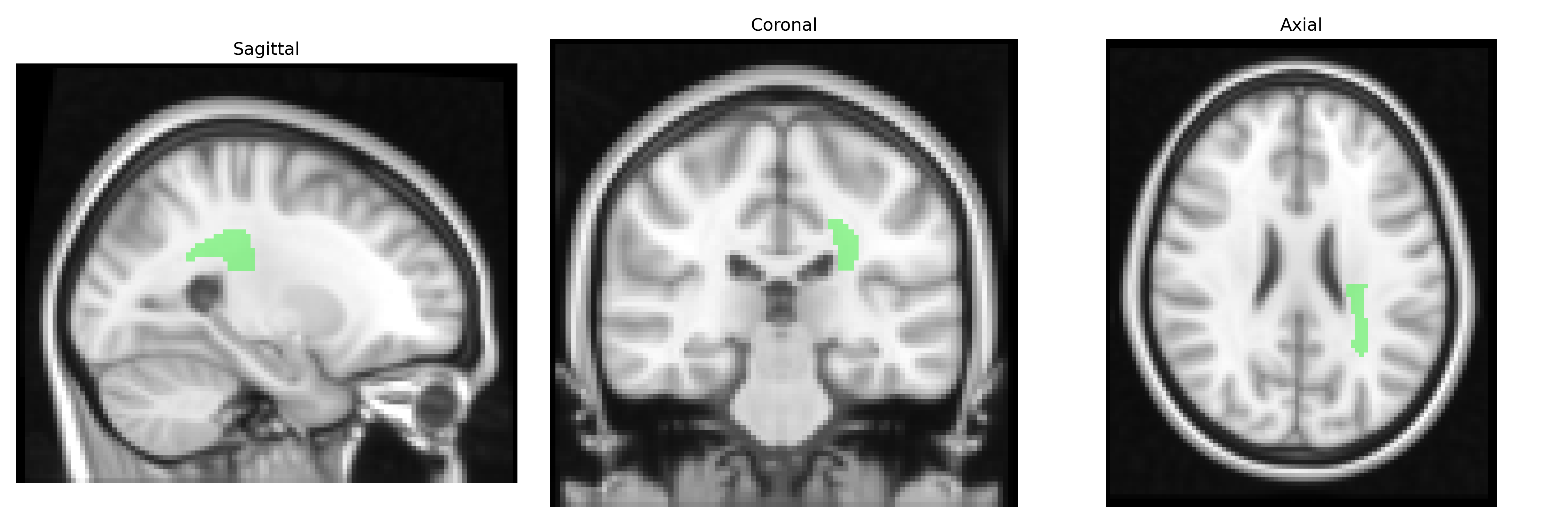
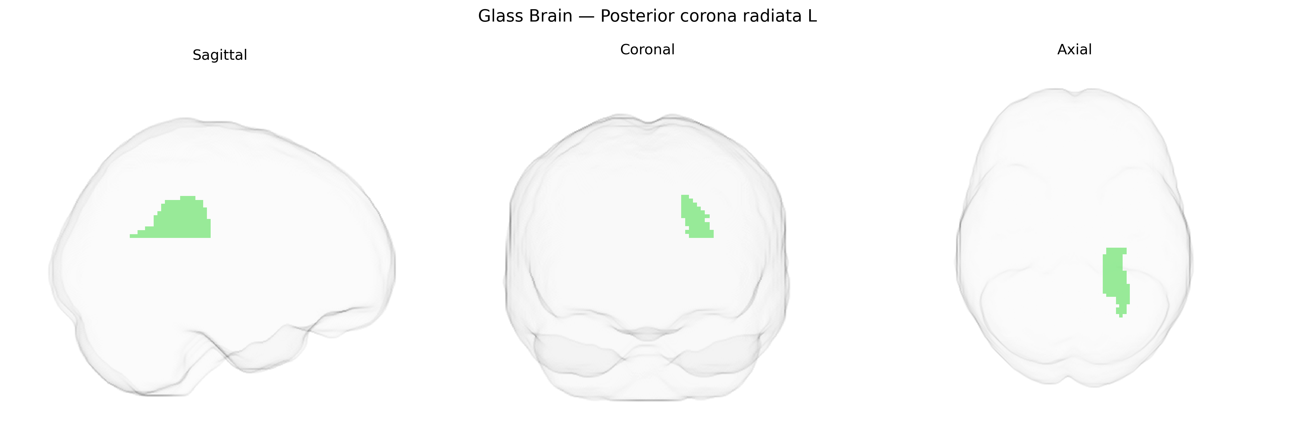
The bilateral posterior corona radiata (left) is a major subcortical white matter tract located superior and posterior to the internal capsule, forming part of the larger corona radiata that fans out between the cerebral cortex and deeper structures. It contains ascending thalamocortical and descending corticospinal and corticopontine fibers connecting posterior parietal, occipital, and some temporal association cortices with the thalamus, brainstem, and spinal cord. Functionally, this region supports sensorimotor integration, visuospatial processing, and higher-order association functions by relaying information between cortical and subcortical networks; lesions in this area can contribute to motor weakness, sensory deficits, and cognitive-visual disturbances depending on the specific fiber bundles involved. There is no direct Wikipedia article for this exact region; a related structure is the Corona radiata (brain).
The bilateral posterior corona radiata, a major white matter tract in the JHU ICBM 2 mm atlas, has been implicated in multiple imaging genetics and GWAS studies that link its microstructural properties—typically measured via diffusion MRI metrics such as fractional anisotropy (FA) and mean diffusivity (MD)—to common genetic variation, particularly in genes involved in myelination, axonal development, and neuroinflammation (e.g., variants in myelin-related genes, oligodendrocyte function pathways, and immune-related loci such as those within the major histocompatibility complex). Large neuroimaging GWAS consortia (such as ENIGMA and UK Biobank-derived studies) have reported associations between posterior corona radiata white matter integrity and polygenic risk scores or specific SNPs related to schizophrenia, bipolar disorder, major depressive disorder, and cognitive performance, suggesting that genetic influences on neurodevelopment and synaptic plasticity partly manifest in this tract. Additionally, white matter measures in the corona radiata have been associated with genetic risk for stroke and small vessel disease, and with loci linked to vascular risk factors (e.g., blood pressure and lipid-related genes), consistent with its vulnerability to ischemic and microvascular pathology. Although findings are typically distributed across the broader corona radiata and neighboring tracts rather than isolated to the posterior segment, these studies collectively indicate that the posterior corona radiata is a genetically sensitive region whose structure and connectivity are shaped by common and polygenic variants tied to psychiatric disorders, cognitive traits, and cerebrovascular disease.
Overview generated by GPT-4o (2026).
Region ID: 28
Hemisphere: bilateral
Atlas: JHU ICBM labels 2mm
Full Quality Version: Download MP4
Full Quality Version: Download MP4
Wali Sidiqyar*, Gaurav Rudravaram*, Elyssa M. McMaster, Trent M. Schwartz, Adam M. Saunders, Kurt G. Schilling, Bennett A. Landman "Introducing SPINS: A Shared Public Visualization Library of Neuroanatomical Structures." Medical Imaging with Deep Learning- short paper
This resource is licensed under CC0 1.0 Universal (Public Domain).