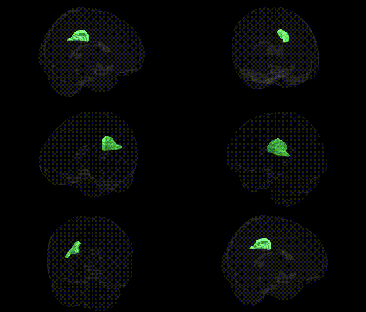
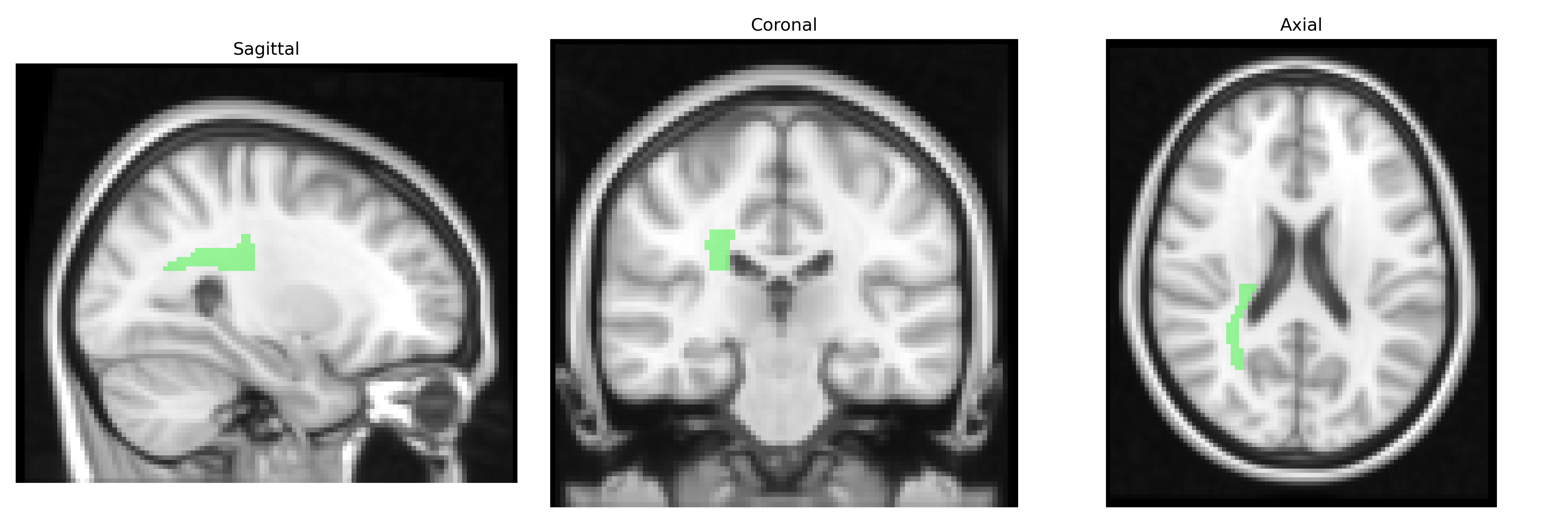
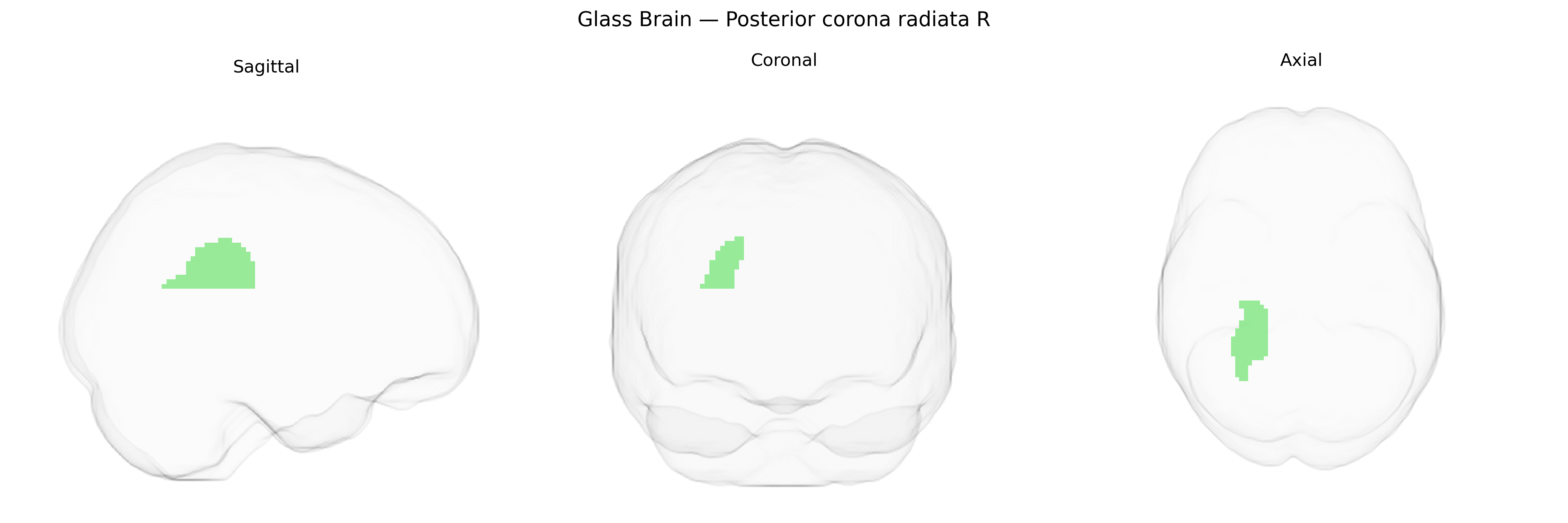
The bilateral Posterior corona radiata (Right) is a major white matter tract segment located in the deep subcortical region of the parietal and occipital lobes, representing the posterior portion of the corona radiata as defined in the JHU ICBM 2 mm atlas. It consists of densely packed projection fibers that fan out between the internal capsule and the cerebral cortex, conveying sensorimotor, visual, and associative information between thalamic nuclei, brainstem structures, and posterior cortical regions, including parietal and occipital association areas. This region plays a key role in higher-order integration of sensory information, visuospatial processing, and coordination of complex cognitive functions dependent on efficient corticothalamic and corticocortical communication. There is no direct link for the posterior corona radiata; a related structure is the Corona radiata (brain).
Genetic associations specific to the bilateral posterior corona radiata (PCR) as defined in the JHU ICBM 2 mm atlas largely come from imaging–genetics and brain-structure GWAS that treat this region as part of broader white-matter tracts in the centrum semiovale and projection fibers to parietal and occipital cortex. Large diffusion MRI GWAS (for example, UK Biobank–based studies of fractional anisotropy and mean diffusivity) have linked common variants in genes involved in axon guidance, myelination, and oligodendrocyte function—such as those in or near NCAN, NTRK3, ROBO1/ROBO2, CNTN4, MAG, and other neurodevelopmental/myelin-related loci—to microstructural measures in corona radiata tracts, with posterior portions often clustering genetically with parietal-occipital networks. Polygenic influences on PCR integrity overlap with risk loci for neuropsychiatric and neurodevelopmental disorders, including schizophrenia, bipolar disorder, major depressive disorder, and autism spectrum disorder, consistent with case–control findings of altered posterior corona radiata white-matter integrity in these conditions; some of these overlaps arise via shared variants in synaptic and calcium-signaling genes (e.g., CACNA1C, GRIN2A) that also affect global or regional white-matter organization. Additional GWAS have tied corona radiata diffusion metrics to cognitive traits (general intelligence, processing speed, educational attainment) and modifiable risk factors such as blood pressure and cardiovascular risk, often mediated by genes influencing small-vessel function and white-matter lesion burden (e.g., loci near COL4A2 and other vascular-ECM genes). Overall, current evidence suggests that the bilateral posterior corona radiata is a genetically influenced component of large-scale frontoparietal and visual–parietal connectivity, with shared polygenic architecture spanning neurodevelopmental patterning, myelination, vascular integrity, and susceptibility to major psychiatric and cognitive phenotypes, though precise locus-level mappings to this specific JHU-defined parcel remain limited and typically inferred from tract- or network-level analyses rather than parcel-specific GWAS.
Overview generated by GPT-4o (2026).
Region ID: 27
Hemisphere: bilateral
Atlas: JHU ICBM labels 2mm
Full Quality Version: Download MP4
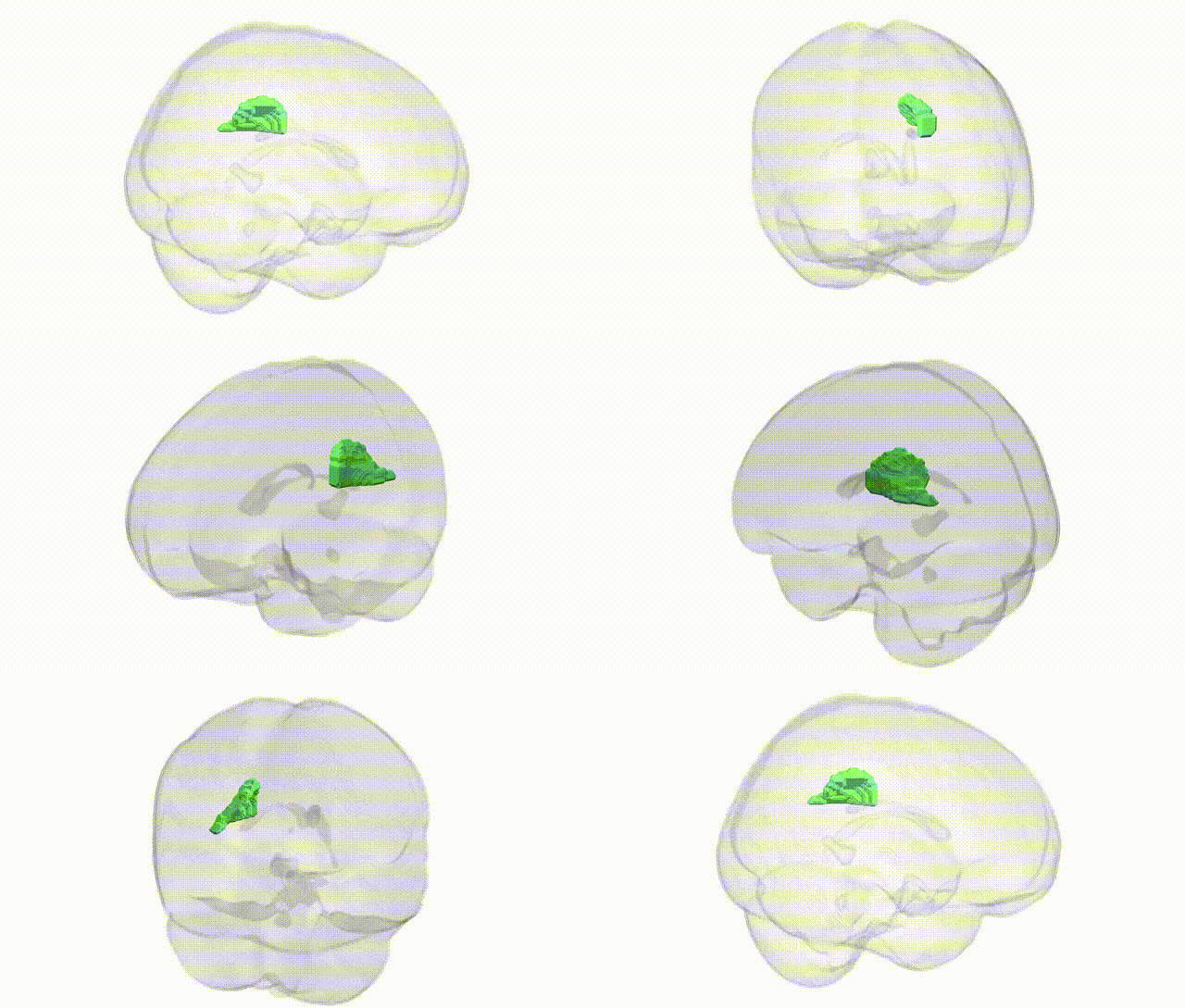
Full Quality Version: Download MP4
Wali Sidiqyar*, Gaurav Rudravaram*, Elyssa M. McMaster, Trent M. Schwartz, Adam M. Saunders, Kurt G. Schilling, Bennett A. Landman "Introducing SPINS: A Shared Public Visualization Library of Neuroanatomical Structures." Medical Imaging with Deep Learning- short paper
This resource is licensed under CC0 1.0 Universal (Public Domain).