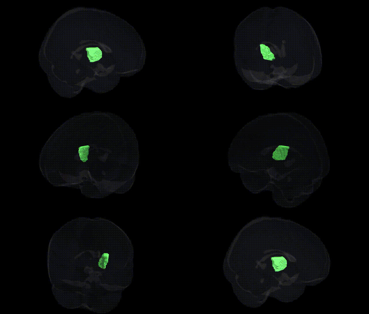
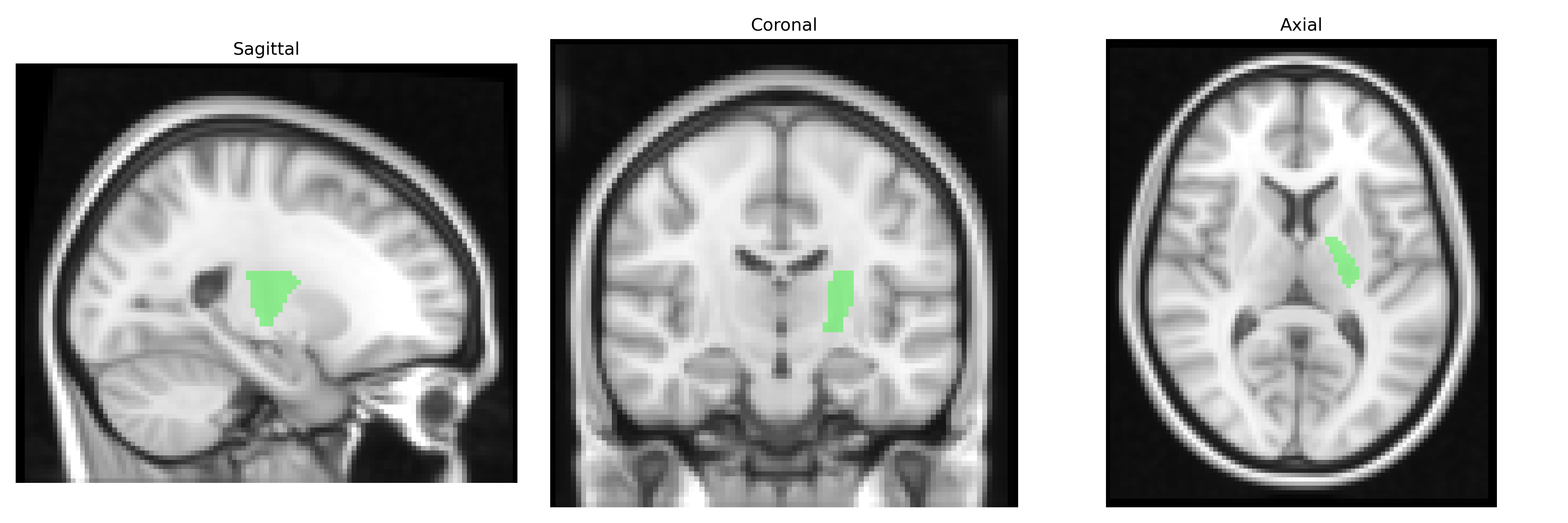
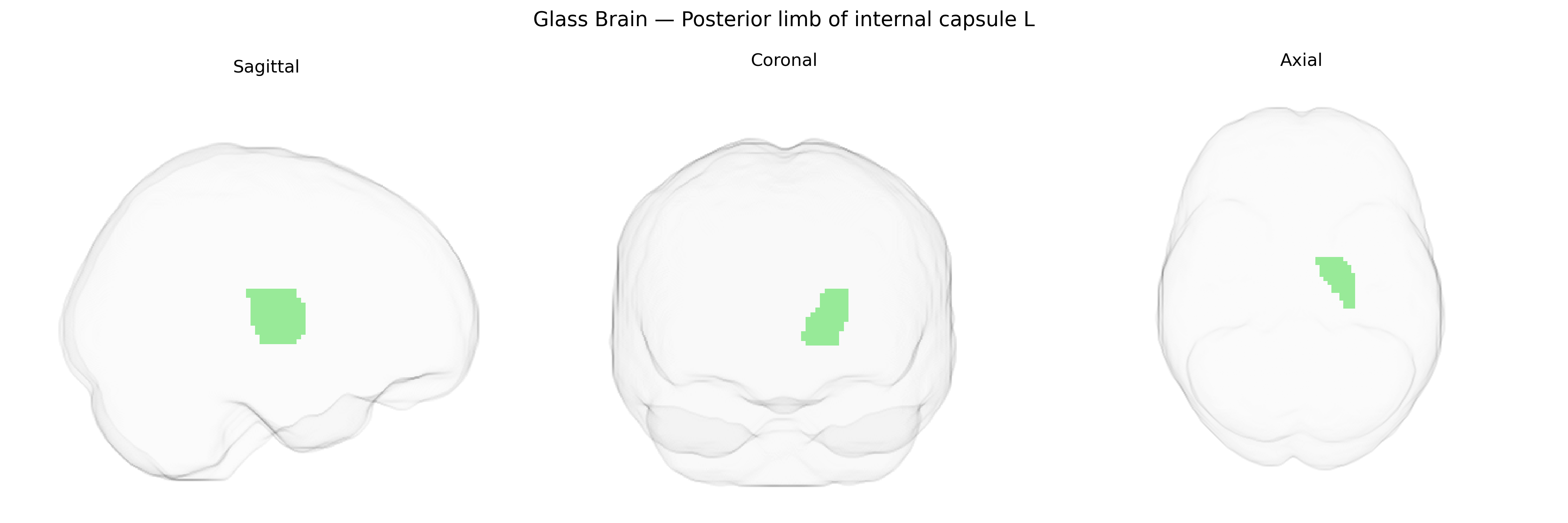
The bilateral Posterior limb of internal capsule L, as defined in the JHU ICBM 2mm white-matter atlas, corresponds to the left-sided portion of the posterior limb of the internal capsule, a major compact white-matter tract situated between the thalamus medially and the lentiform nucleus laterally. It contains densely packed ascending and descending fibers, including corticospinal, corticobulbar, and thalamocortical projections, which are crucial for voluntary motor control, somatosensory transmission from the body, and integration of thalamic input to the cerebral cortex. Lesions in this region commonly produce contralateral motor deficits and sensory disturbances due to the concentration of projection fibers. There is no dedicated Wikipedia entry specifically for the “posterior limb of internal capsule,” but it is a subregion of the Internal capsule.
The bilateral posterior limb of the internal capsule (PLIC), as defined in the JHU ICBM labels 2 mm atlas, is a major white matter tract carrying corticospinal and thalamocortical fibers, and genetic associations to this region arise primarily from imaging genetics and GWAS of white matter microstructure rather than region-specific case studies. Large-scale diffusion MRI GWAS (such as UK Biobank-based studies) have identified multiple loci influencing fractional anisotropy and mean diffusivity in internal capsule tracts, including variants near genes involved in axon guidance, myelination, and neurodevelopment (for example, loci near CNTN4, NTRK3, and genes in the netrin and semaphorin pathways), and these microstructural measures in the internal capsule show heritability estimates often in the 30–60% range. Polygenic influences related to neuropsychiatric disorders (schizophrenia, bipolar disorder, major depression), neurodevelopmental conditions (ADHD, autism spectrum disorder), and cognitive traits (general intelligence, processing speed) have been linked to altered integrity of the PLIC, with some studies demonstrating overlap between disease-associated genetic risk scores and internal capsule diffusion metrics. In addition, GWAS of stroke and small vessel disease have implicated loci (e.g., near COL4A1/COL4A2, HDAC9) that relate to vulnerability of internal capsule fibers, consistent with the PLIC’s role as a common site of lacunar infarcts; however, these findings generally reflect tract-level or global white matter associations, rather than genetic effects uniquely specific to the left PLIC label in the JHU atlas.
Overview generated by GPT-4o (2026).
Region ID: 20
Hemisphere: bilateral
Atlas: JHU ICBM labels 2mm
Full Quality Version: Download MP4
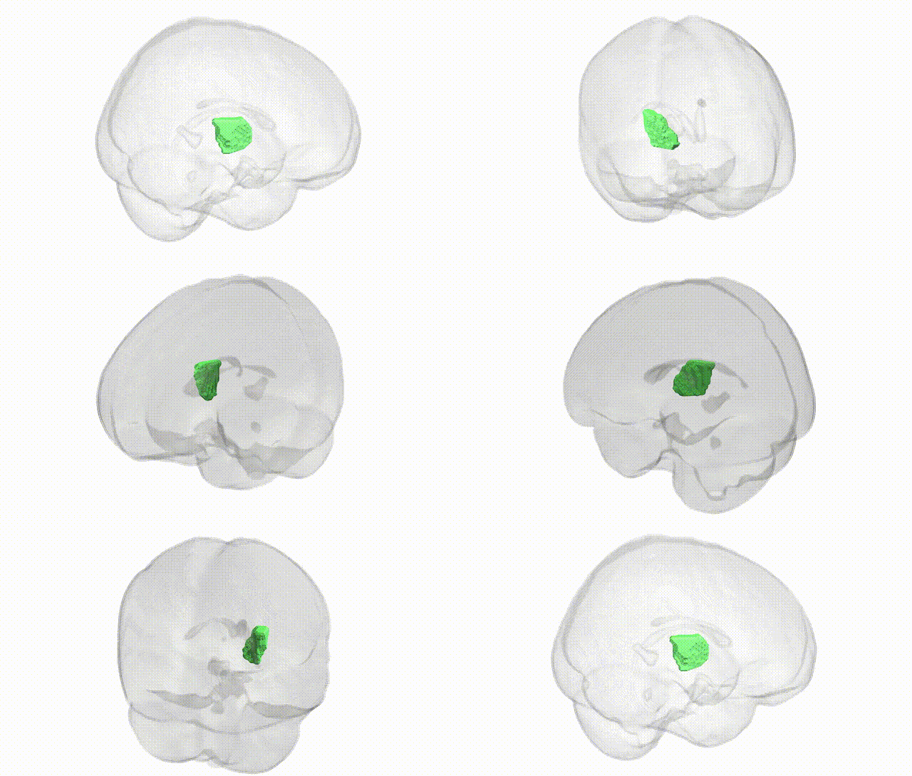
Full Quality Version: Download MP4
Wali Sidiqyar*, Gaurav Rudravaram*, Elyssa M. McMaster, Trent M. Schwartz, Adam M. Saunders, Kurt G. Schilling, Bennett A. Landman "Introducing SPINS: A Shared Public Visualization Library of Neuroanatomical Structures." Medical Imaging with Deep Learning- short paper
This resource is licensed under CC0 1.0 Universal (Public Domain).