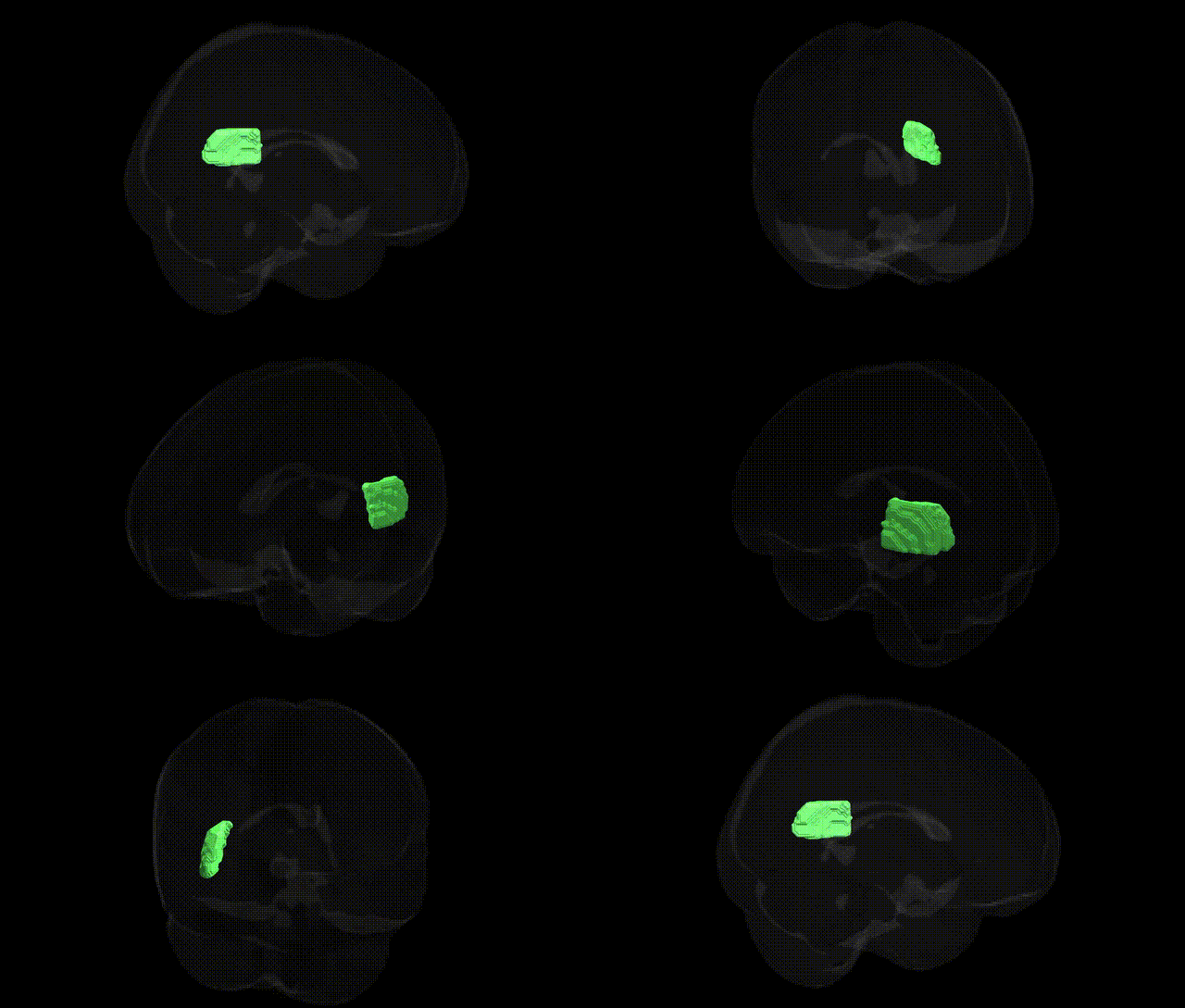
The bilateral Posterior thalamic radiation (include optic radiation) R, as defined in the JHU ICBM 2mm Atlas, is a major white matter pathway connecting the posterior and lateral thalamic nuclei with occipital and posterior temporal cortical areas, including the primary and associative visual cortices. This tract encompasses the optic radiation, a critical component of the visual system that conveys visual information from the lateral geniculate nucleus to the striate cortex (V1), and also includes adjacent posterior thalamocortical fibers participating in multimodal sensory integration. Structurally, it courses through the posterior limb and retrolenticular part of the internal capsule, forming a key part of the subcortical–cortical communication network for visual perception, visuospatial processing, and higher-order visual cognition. Damage to this region is frequently associated with visual field deficits (such as homonymous hemianopia) and may contribute to broader impairments in sensory processing and cortico-thalamic signaling.
The bilateral posterior thalamic radiation, including the optic radiation, has been implicated in several genetic and GWAS-based findings primarily through diffusion MRI studies of white matter microstructure and imaging–genetics analyses. Variants in genes involved in axonal guidance, myelination, and synaptic function (e.g., NTRK1/3, CNTNAP2, MAG, and contactin-related loci) have been associated with fractional anisotropy and other diffusion metrics in this tract, reflecting genetic control of thalamo-occipital connectivity and visual pathway integrity. Large-scale GWAS of brain imaging phenotypes (such as those from UK Biobank and ENIGMA) have identified multiple loci influencing the microstructure of posterior thalamic/optic radiations, with some overlapping genetic risk for schizophrenia, major depressive disorder, bipolar disorder, and autism spectrum disorder, suggesting that genetically driven alterations in this region may contribute to visual processing, attentional, and cognitive deficits observed in these conditions. Additionally, variants in genes related to neurodevelopment and neurodegeneration (including APOE and other Alzheimer’s disease–associated loci) have been linked to white matter changes encompassing the posterior thalamic radiation, connecting genetic risk for dementia and cognitive decline to structural disruption of this pathway. GWAS of visual function and visual acuity have also pointed to shared genetic influences on optic radiation microstructure, highlighting this tract as a convergent substrate where genetic variation impacts both clinical disorders and intermediate imaging-based endophenotypes.
Overview generated by GPT-4o (2026).
Region ID: 29
Hemisphere: bilateral
Atlas: JHU ICBM labels 2mm
Full Quality Version: Download MP4
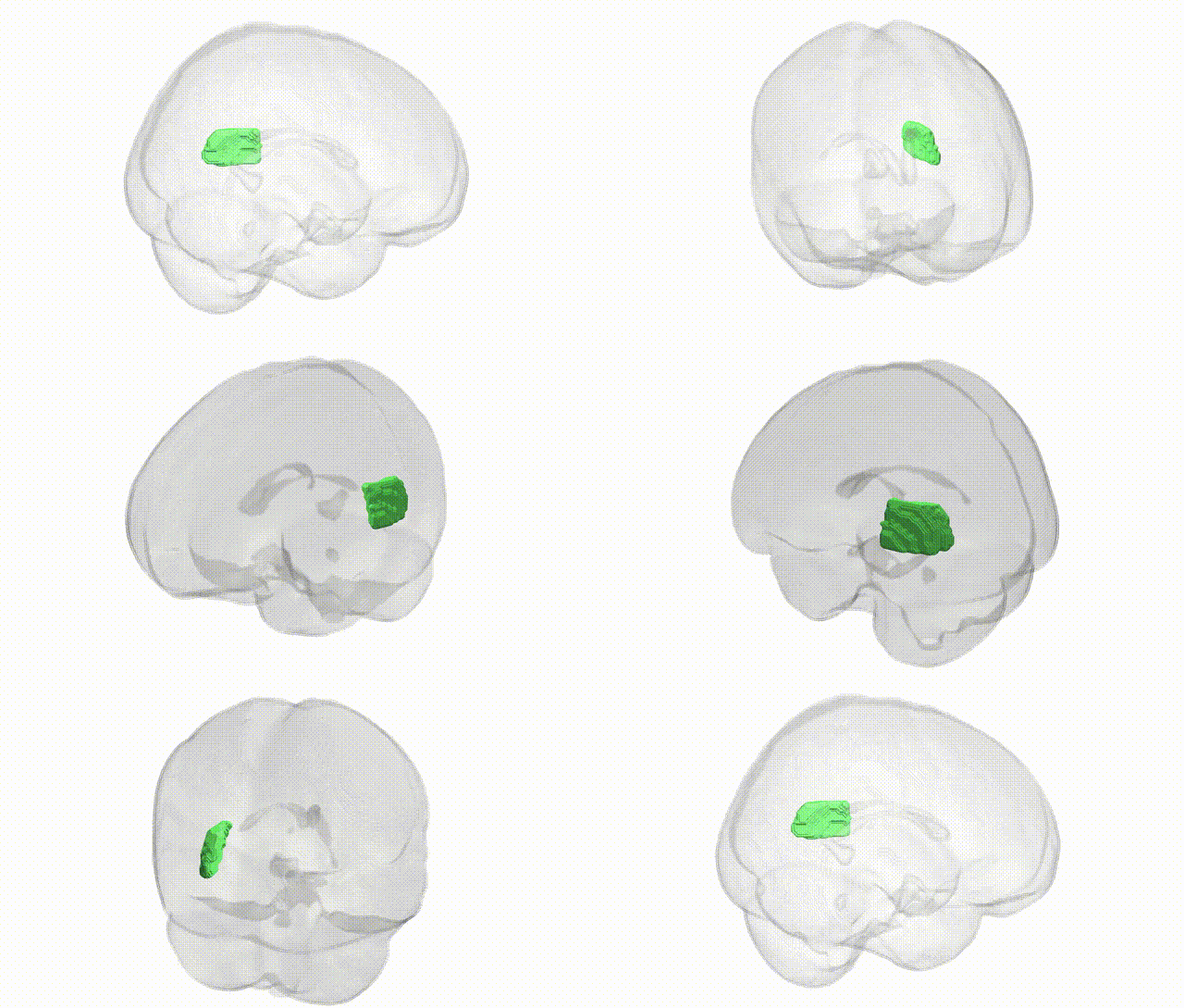
Full Quality Version: Download MP4
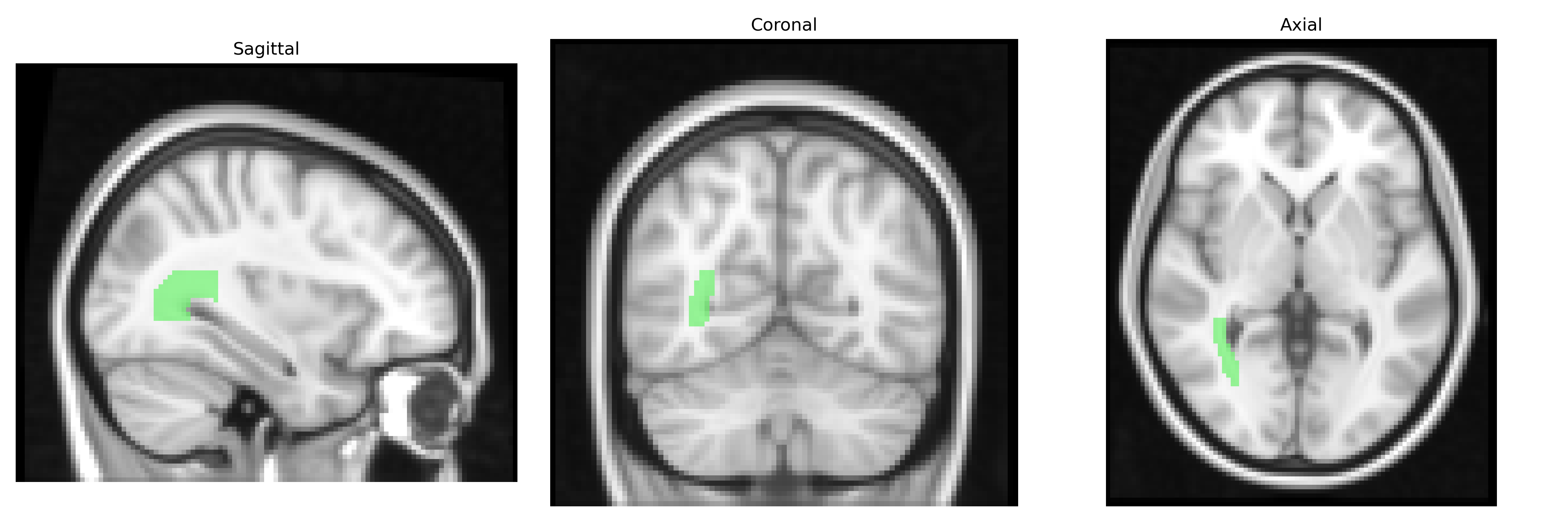
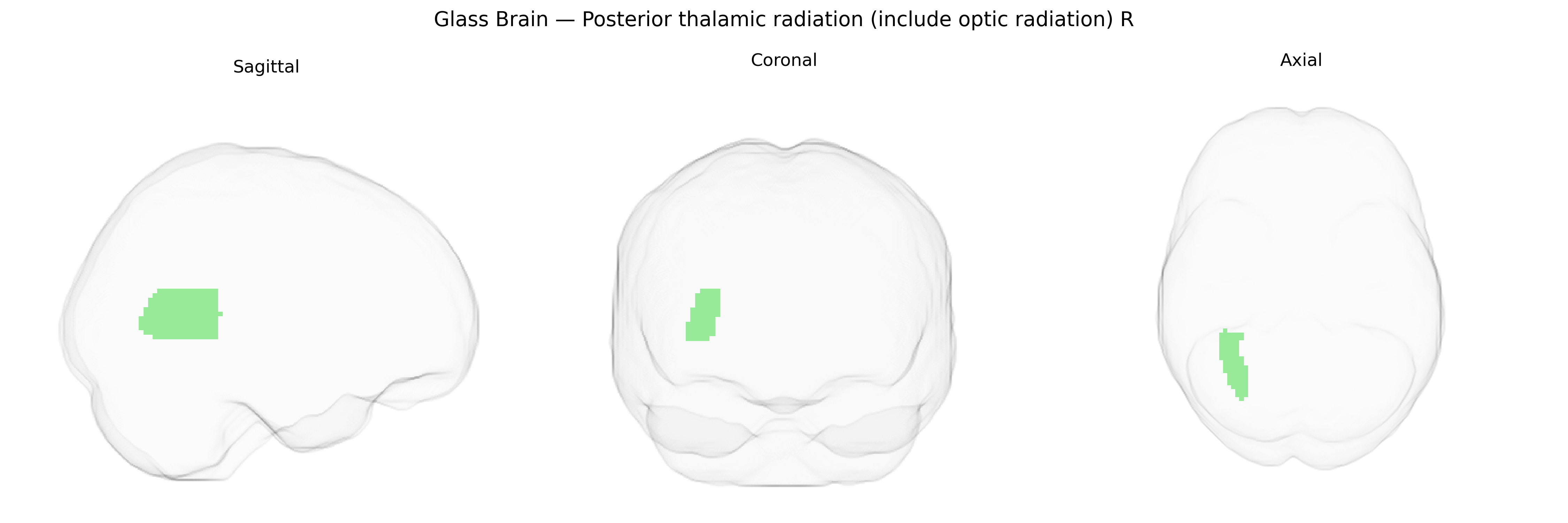
Wali Sidiqyar*, Gaurav Rudravaram*, Elyssa M. McMaster, Trent M. Schwartz, Adam M. Saunders, Kurt G. Schilling, Bennett A. Landman "Introducing SPINS: A Shared Public Visualization Library of Neuroanatomical Structures." Medical Imaging with Deep Learning- short paper
This resource is licensed under CC0 1.0 Universal (Public Domain).