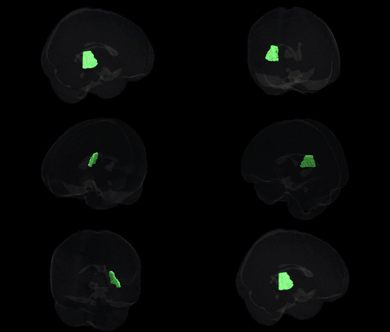
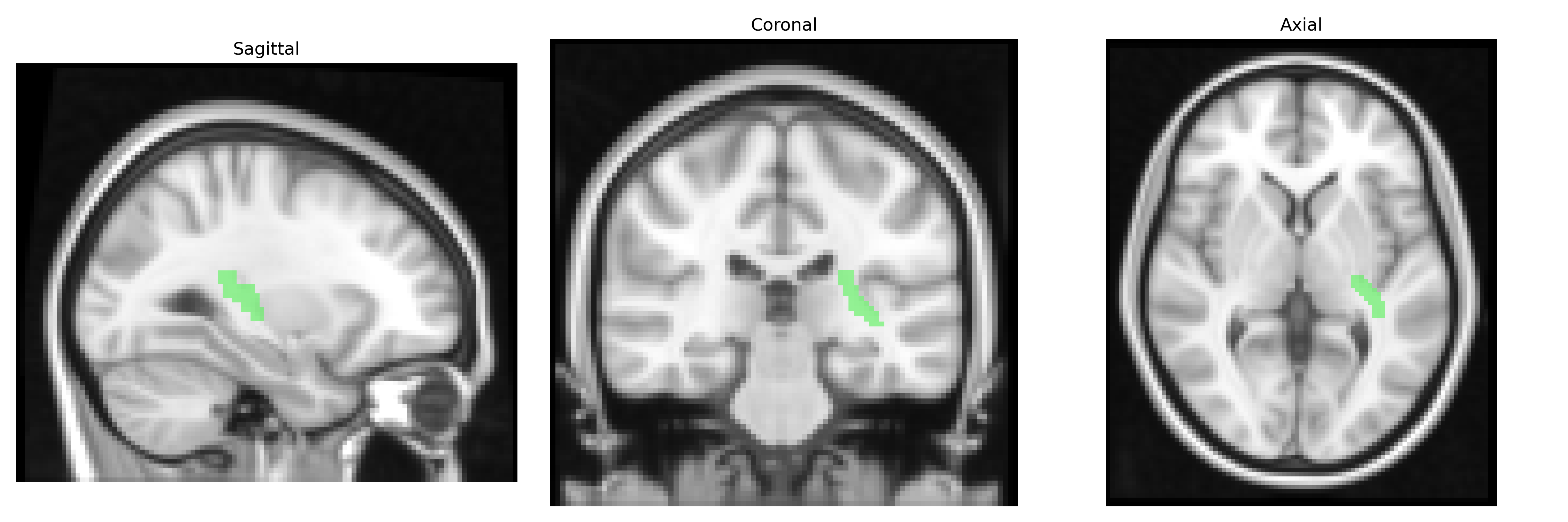
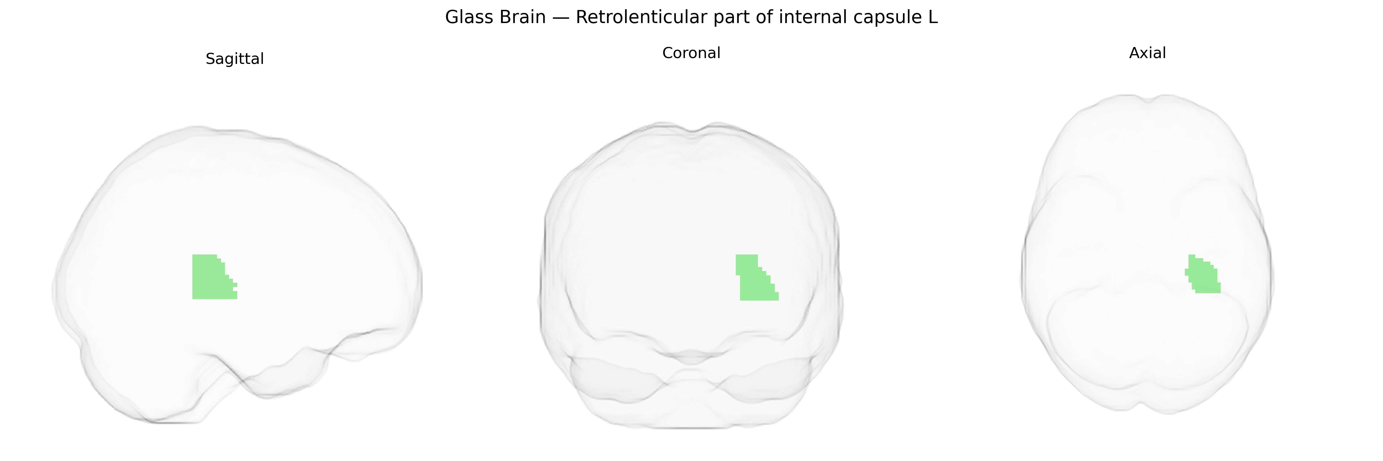
The bilateral retrolenticular part of the internal capsule is a posterior segment of the internal capsule that lies behind the lentiform nucleus and contains major projection fiber pathways, including portions of the optic radiation and posterior thalamic radiations, as well as corticopontine and other cortico-subcortical tracts. Functionally, it is critical for visual processing and visuospatial integration via connections between the lateral geniculate nucleus and the occipital cortex, and it also participates in sensorimotor and associative information transfer between cortex and thalamus. Lesions in this region can produce visual field deficits, particularly homonymous hemianopia, along with possible disturbances in higher-order perceptual and cognitive functions due to disruption of thalamocortical connectivity. There is no direct link; a closely related structure is the Internal capsule.
The bilateral retrolenticular part of the internal capsule, as defined in the JHU ICBM labels 2mm atlas, has been implicated in several imaging genetics and GWAS studies through its role in major white‑matter tracts linking occipital, temporal, and parietal cortices with subcortical structures. Variants in genes associated with myelination, axon guidance, and neurodevelopment (e.g., NTRK1/3, MAG, CNTN4, and other loci near neurodevelopmental and oligodendrocyte-related genes) have been reported to influence fractional anisotropy and other diffusion MRI metrics in this region or overlapping tracts such as the optic radiations and posterior limb of the internal capsule. Large-scale GWAS of white-matter microstructure (UK Biobank and related cohorts) have identified multiple genome-wide significant loci affecting diffusion properties in internal capsule and retrolenticular pathways, some of which colocalize with risk loci for schizophrenia, bipolar disorder, major depressive disorder, and cognitive traits including general intelligence and processing speed. Imaging genetics studies in multiple sclerosis, small-vessel cerebrovascular disease, and ischemic stroke have further linked polygenic risk scores and specific susceptibility loci (e.g., near COL4A1/2, ZIC4, and other vascular or inflammatory genes) with structural integrity and lesion burden in this posterior internal capsule region, consistent with its vulnerability to demyelination and lacunar infarcts. In addition, GWAS of neurodevelopmental and neuropsychiatric conditions such as autism spectrum disorder and ADHD, while not always targeting this region explicitly, have shown that risk variants affecting large-scale fronto-parietal and visual pathways also modulate microstructural measures and connectivity passing through the retrolenticular internal capsule, supporting a shared genetic architecture between white-matter organization in this tract and a broad range of cognitive and psychiatric phenotypes.
Overview generated by GPT-4o (2026).
Region ID: 22
Hemisphere: bilateral
Atlas: JHU ICBM labels 2mm
Full Quality Version: Download MP4
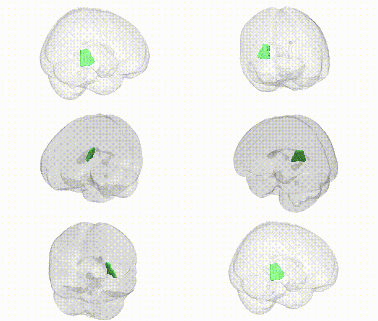
Full Quality Version: Download MP4
Wali Sidiqyar*, Gaurav Rudravaram*, Elyssa M. McMaster, Trent M. Schwartz, Adam M. Saunders, Kurt G. Schilling, Bennett A. Landman "Introducing SPINS: A Shared Public Visualization Library of Neuroanatomical Structures." Medical Imaging with Deep Learning- short paper
This resource is licensed under CC0 1.0 Universal (Public Domain).