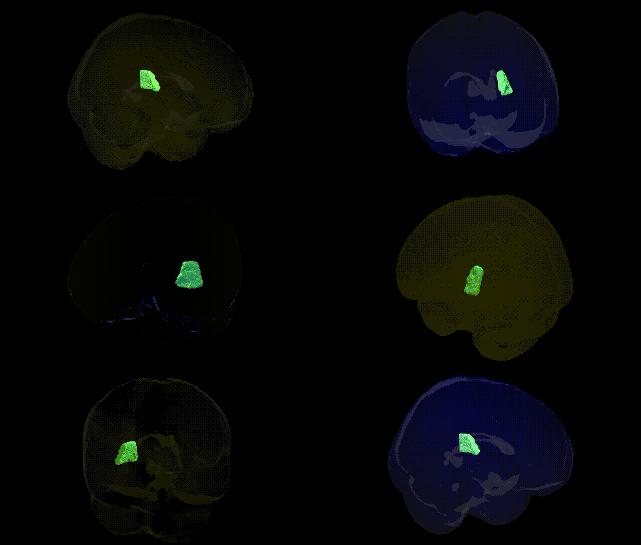
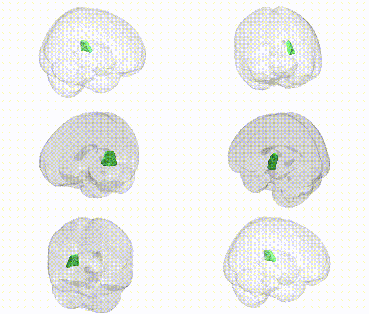
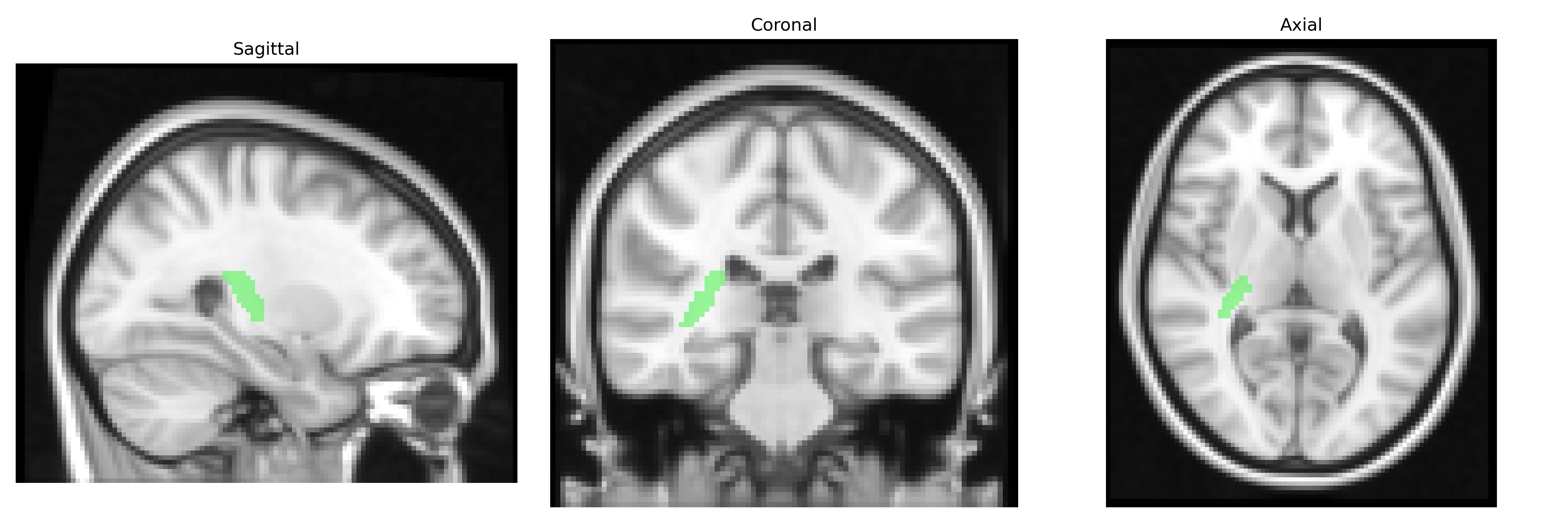
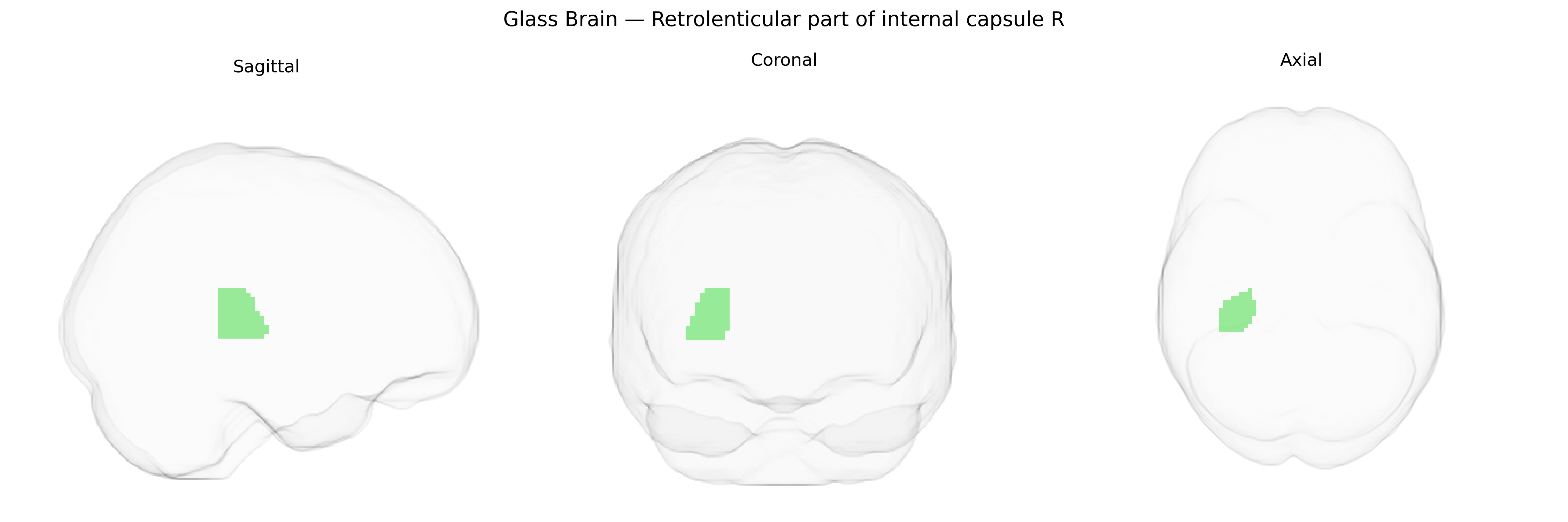
The bilateral retrolenticular part of the internal capsule (right) is a segment of the posterior internal capsule situated behind the lentiform nucleus, containing major projection fibers that connect thalamic nuclei with occipital and posterior parietal cortices. It carries a substantial portion of the optic radiations from the lateral geniculate nucleus to the primary visual cortex, as well as components of the auditory radiations and other posterior thalamocortical pathways involved in sensory integration and visuospatial processing. Damage to this region can lead to contralateral visual field deficits (such as homonymous hemianopia) and may contribute to higher-order perceptual and cognitive impairments depending on the specific fibers involved. There is no direct Wikipedia article for this exact subdivision; a related structure is the Internal capsule.
The bilateral retrolenticular part of the internal capsule (RLIC) as defined in the JHU ICBM 2 mm atlas is a major white matter pathway carrying posterior thalamic radiations and optic radiations, and has been implicated in multiple imaging genetics and GWAS studies of white matter microstructure. Diffusion MRI GWAS consortia such as ENIGMA and UK Biobank have reported significant heritability of fractional anisotropy (FA) and mean diffusivity (MD) in the RLIC, with associations involving common variants in genes related to axon guidance, myelination, and neural development, including loci near or within genes such as DRD2, BDNF, CNTN4, and MAG, although specific effect sizes are modest and often shared across multiple tracts rather than unique to the RLIC. Polygenic risk for schizophrenia and bipolar disorder has been linked to reduced FA in posterior internal capsule and retrolenticular fibers, suggesting that distributed psychiatric risk alleles contribute to microstructural changes in this region, and similar polygenic influences have been reported for major depressive disorder and autism spectrum disorder in large-scale imaging genetics analyses. In vascular and neurodegenerative conditions, risk variants for cerebral small vessel disease, hypertension, and Alzheimer’s disease (e.g., in APOE and COL4A1/2 pathways) have been associated with higher white matter hyperintensity burden and microstructural disruption involving the internal capsule, including its retrolenticular portion. Additionally, GWAS of brain connectivity and structural network organization have implicated common genetic variants that modulate thalamocortical and visual pathway integrity, with the RLIC frequently appearing as a key tract through which these genetically influenced network changes are expressed, though causal, tract-specific genetic effects remain incompletely resolved.
Overview generated by GPT-4o (2026).
Region ID: 21
Hemisphere: bilateral
Atlas: JHU ICBM labels 2mm
Full Quality Version: Download MP4
Full Quality Version: Download MP4
Wali Sidiqyar*, Gaurav Rudravaram*, Elyssa M. McMaster, Trent M. Schwartz, Adam M. Saunders, Kurt G. Schilling, Bennett A. Landman "Introducing SPINS: A Shared Public Visualization Library of Neuroanatomical Structures." Medical Imaging with Deep Learning- short paper
This resource is licensed under CC0 1.0 Universal (Public Domain).