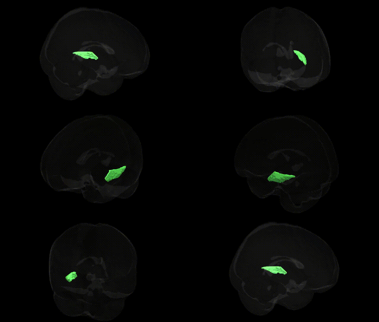
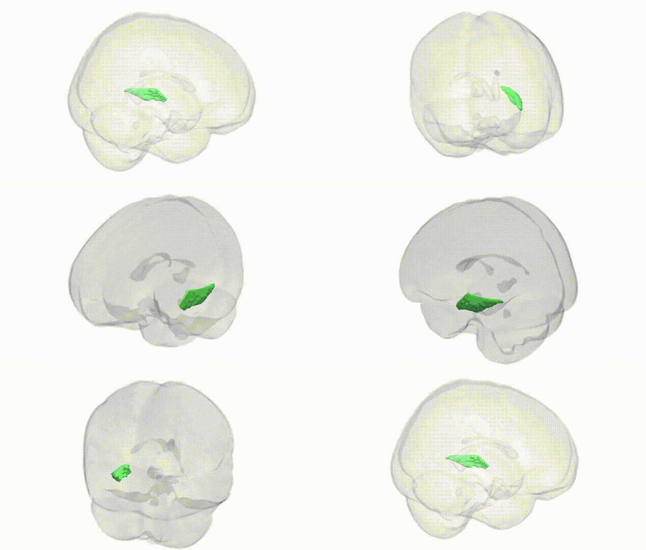
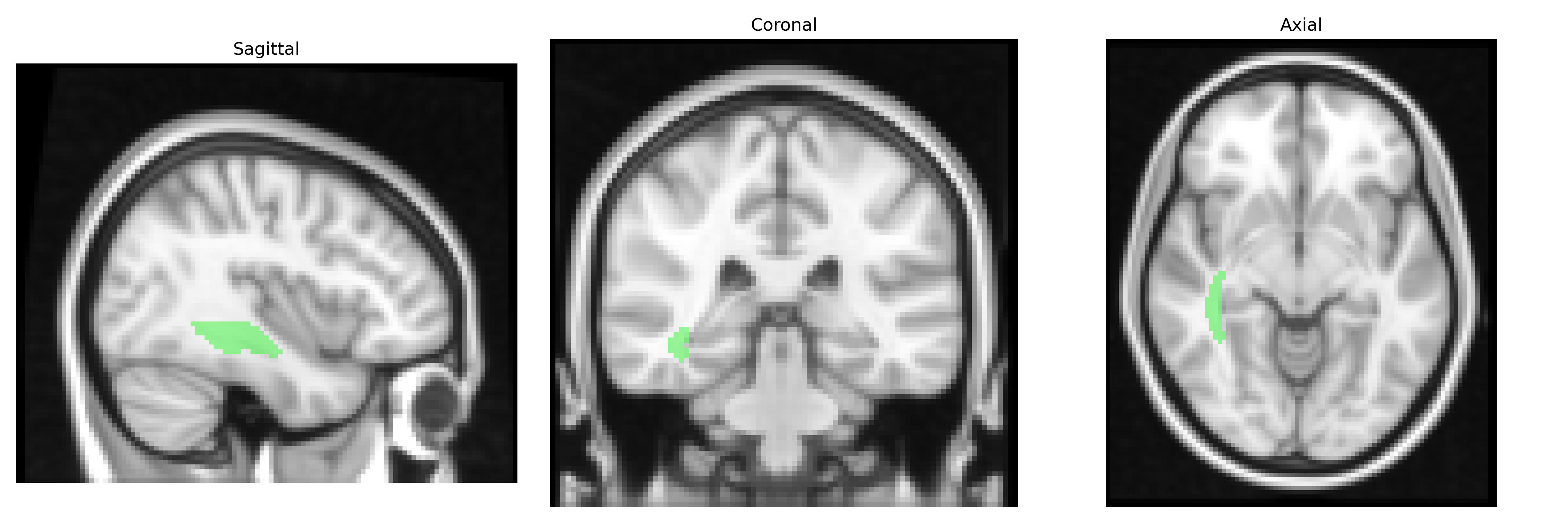
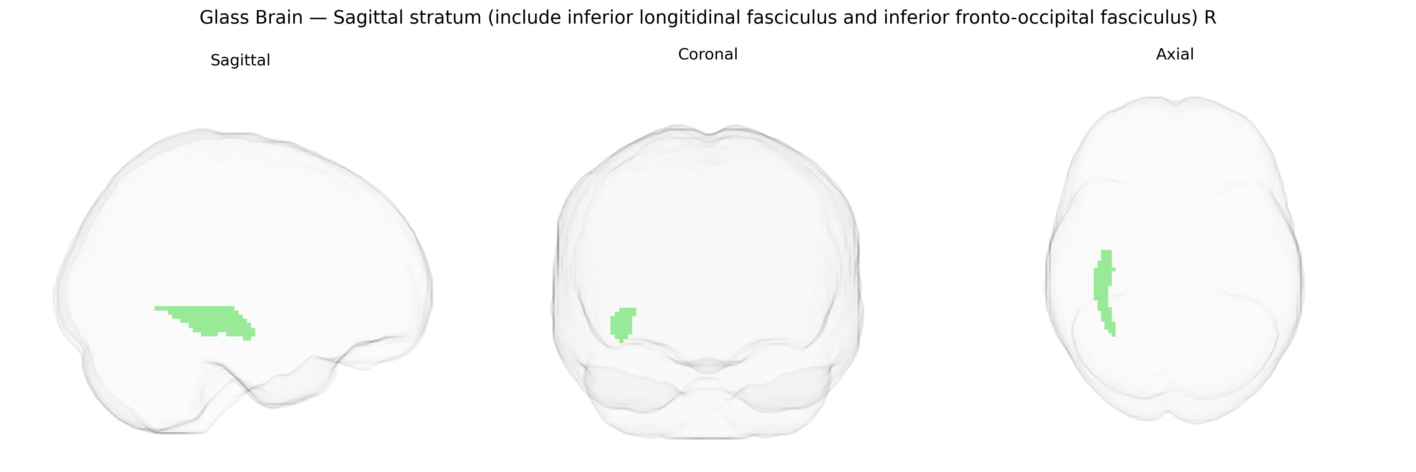
The bilateral sagittal stratum (including the inferior longitudinal fasciculus and inferior fronto-occipital fasciculus) is a major deep white matter sheet located in the posterior cerebral hemispheres, running parallel to the ventricular system and underlying the temporal and occipital lobes. It comprises long association fibers that connect occipital visual regions with temporal cortex (via the inferior longitudinal fasciculus) and with frontal regions (via the inferior fronto-occipital fasciculus), supporting visual object recognition, semantic processing, and integration of visual information with higher-order cognitive and language functions. The sagittal stratum lies lateral to the atrium of the lateral ventricle and medial to the temporal and occipital cortices, forming part of the broader ventral visual and semantic pathways and interacting with limbic and association networks involved in memory and perception. There is no direct Wikipedia article for the sagittal stratum; see the related Inferior fronto-occipital fasciculus and Inferior longitudinal fasciculus.
The bilateral sagittal stratum (including the inferior longitudinal fasciculus and inferior fronto‑occipital fasciculus as defined in the JHU ICBM 2 mm atlas) has been implicated in multiple imaging–genetics and GWAS-based neuroimaging studies, which commonly use diffusion MRI measures such as fractional anisotropy (FA) or mean diffusivity as endophenotypes. Variants in genes related to axonal guidance, myelination, and oligodendrocyte function (for example, NTRK1/2, MAG, and other myelin-related loci) have shown associations with microstructural properties of these tracts in large consortia such as ENIGMA-DTI and UK Biobank imaging GWAS, though single specific genes often explain only a small fraction of variance. Polygenic risk scores for schizophrenia, bipolar disorder, major depressive disorder, and autism spectrum disorder have been associated with altered FA or structural connectivity in the inferior longitudinal and inferior fronto‑occipital fasciculi, suggesting that distributed common genetic risk for psychiatric illness partly manifests as microstructural changes in these pathways. Additionally, GWAS of cognitive performance, general intelligence, reading ability, and educational attainment have linked common variants in multiple loci (e.g., on chromosomes 3p, 6p, and 17q in various studies) with differences in white matter integrity in temporo‑occipital association tracts, including the inferior longitudinal fasciculus, while neurodegeneration-related loci (e.g., APOE and other Alzheimer’s disease risk genes) have been associated with reduced integrity in posterior association fibers that overlap the sagittal stratum. Overall, genetic associations with this region are highly polygenic and distributed, with no single locus uniquely defining risk, but with convergent evidence that genes involved in neurodevelopment, synaptic function, and myelination contribute to inter-individual variation in its microstructure and to vulnerability for psychiatric and neurodegenerative disorders.
Overview generated by GPT-4o (2026).
Region ID: 31
Hemisphere: bilateral
Atlas: JHU ICBM labels 2mm
Full Quality Version: Download MP4
Full Quality Version: Download MP4
Wali Sidiqyar*, Gaurav Rudravaram*, Elyssa M. McMaster, Trent M. Schwartz, Adam M. Saunders, Kurt G. Schilling, Bennett A. Landman "Introducing SPINS: A Shared Public Visualization Library of Neuroanatomical Structures." Medical Imaging with Deep Learning- short paper
This resource is licensed under CC0 1.0 Universal (Public Domain).