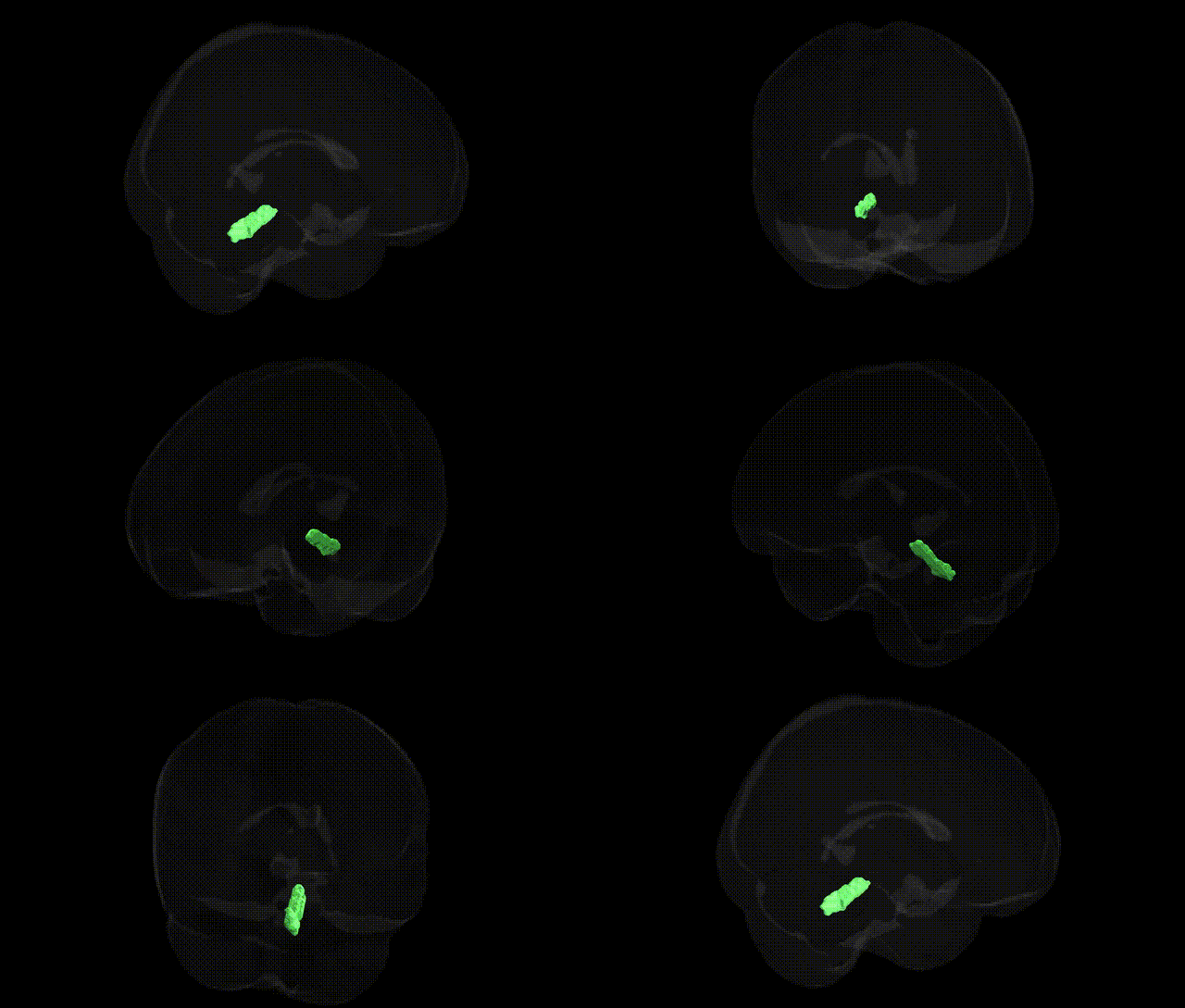
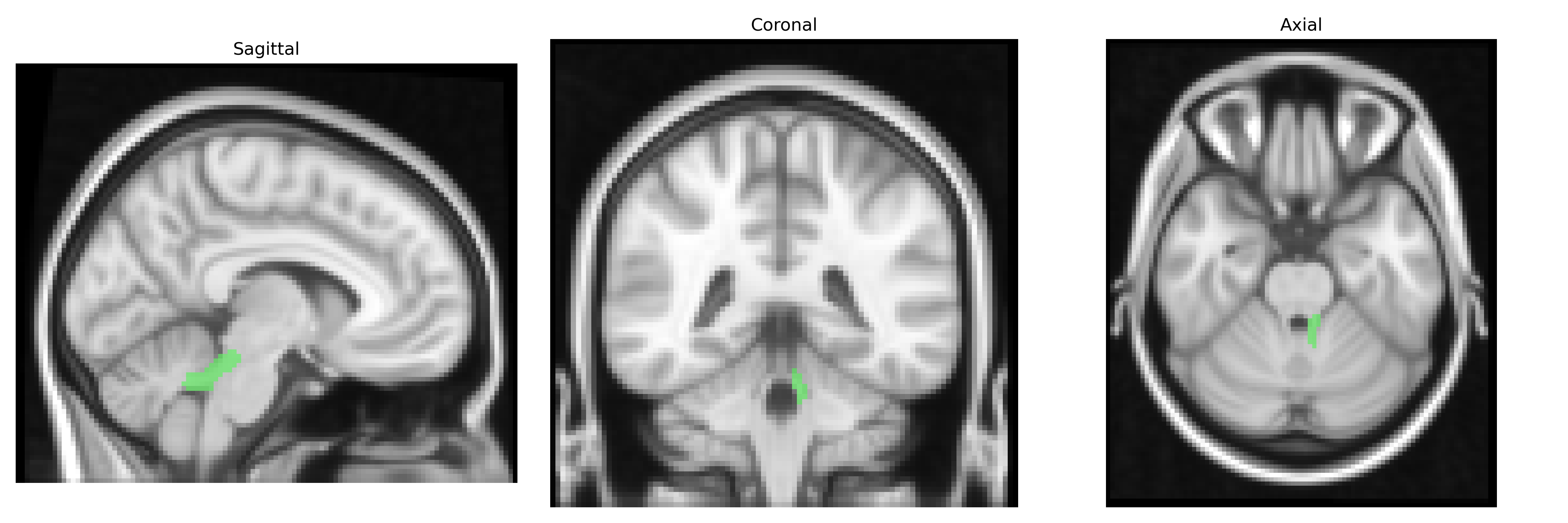
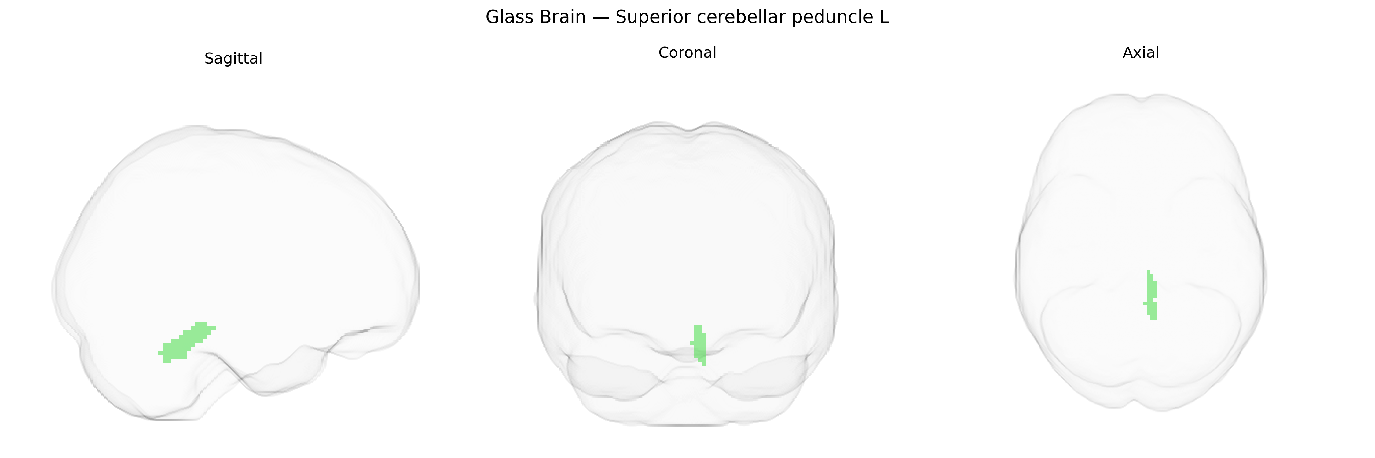
The bilateral Superior cerebellar peduncle L, as defined in the JHU ICBM 2 mm atlas, corresponds to the left superior cerebellar peduncle, a major efferent (and partially afferent) white matter tract connecting the cerebellum to the midbrain. It primarily carries output from deep cerebellar nuclei—especially the dentate nucleus—to the red nucleus and thalamus, thereby influencing motor planning, coordination, and fine-tuning of voluntary movements, as well as aspects of cognitive and oculomotor control. Anatomically, it courses anterosuperiorly from the cerebellar hemispheres and vermis toward the dorsal aspect of the brainstem, forming an essential component of cerebello-thalamo-cortical and cerebello-rubral circuits implicated in motor learning and error correction. Superior cerebellar peduncle
The bilateral superior cerebellar peduncle, labeled in the JHU ICBM 2 mm atlas as tract L (left) and its right counterpart, functions as a major efferent pathway linking the cerebellum to midbrain and thalamic structures, and genetic associations typically emerge from imaging‑genetics and GWAS of diffusion MRI measures such as fractional anisotropy or tract integrity. Large neuroimaging GWAS consortia (e.g., ENIGMA, UK Biobank) have identified common variants influencing microstructural properties in this tract, often near genes implicated in axon guidance, myelination, and neurodevelopment, including loci in or near genes such as CNTN4, NTRK3, and various myelin‑related genes, though findings are generally polygenic rather than dominated by single large‑effect variants. Altered superior cerebellar peduncle structure has been linked, via imaging‑genetics designs, to neurodevelopmental and psychiatric conditions such as autism spectrum disorder, schizophrenia, and ADHD, as well as movement and coordination disorders, with some studies reporting that risk alleles for these conditions partially exert their effects through cerebellar white‑matter pathways; rare variants and Mendelian mutations affecting cerebellar development (e.g., in Joubert syndrome–related genes like AHI1 and CEP290) often show structural abnormalities involving the superior cerebellar peduncles, although these are typically described at the level of cerebellar and brainstem malformations rather than as isolated tract findings. Overall, current evidence supports a diffuse, polygenic influence of neurodevelopmental and myelination‑related genes on the microstructure of the superior cerebellar peduncles, with downstream relevance for cognitive, motor, and psychiatric phenotypes, but no single, robust, tract‑specific GWAS locus has yet achieved the status of a well‑replicated, clinically actionable genetic marker for this region.
Overview generated by GPT-4o (2026).
Region ID: 14
Hemisphere: bilateral
Atlas: JHU ICBM labels 2mm
Full Quality Version: Download MP4
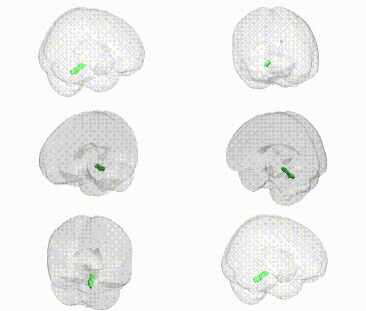
Full Quality Version: Download MP4
Wali Sidiqyar*, Gaurav Rudravaram*, Elyssa M. McMaster, Trent M. Schwartz, Adam M. Saunders, Kurt G. Schilling, Bennett A. Landman "Introducing SPINS: A Shared Public Visualization Library of Neuroanatomical Structures." Medical Imaging with Deep Learning- short paper
This resource is licensed under CC0 1.0 Universal (Public Domain).