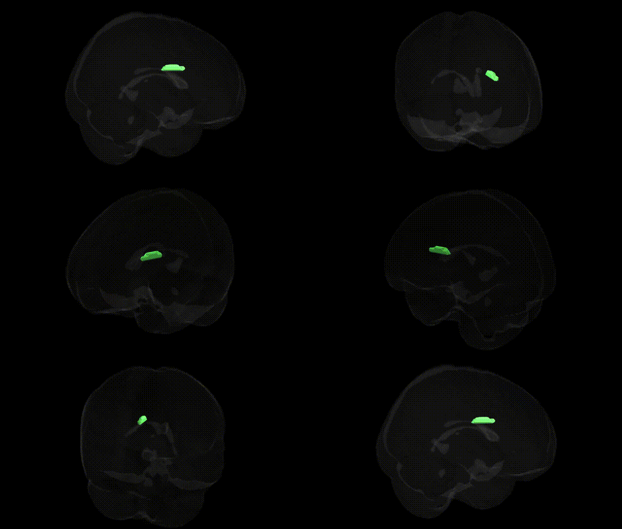
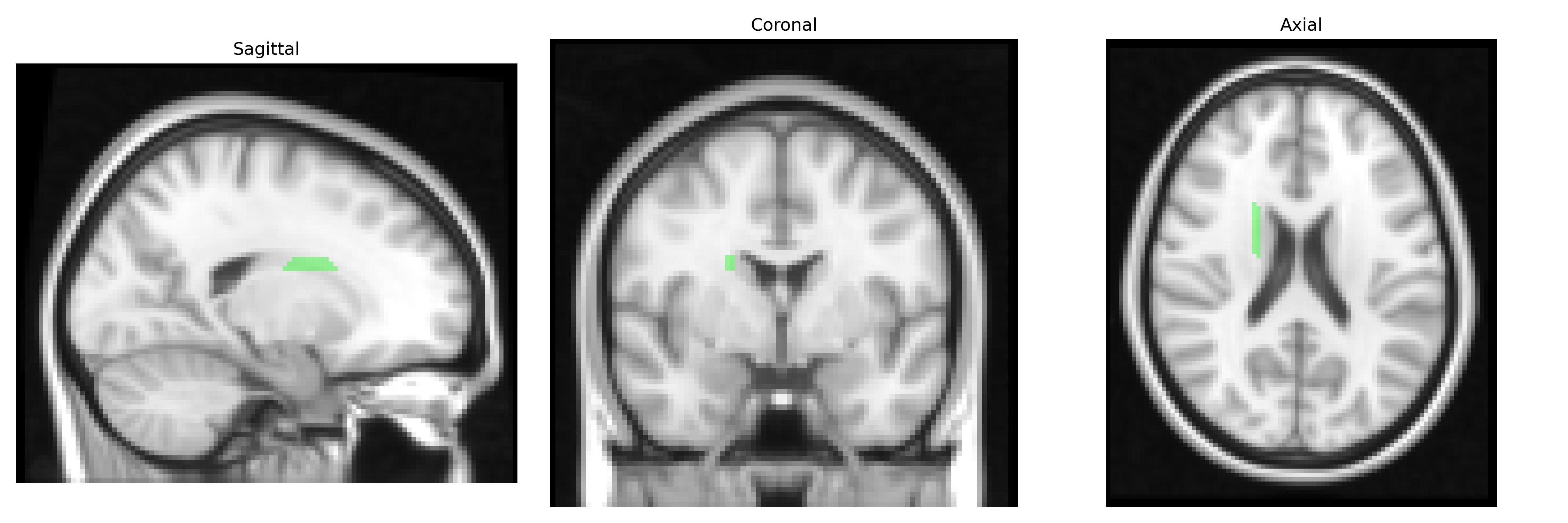
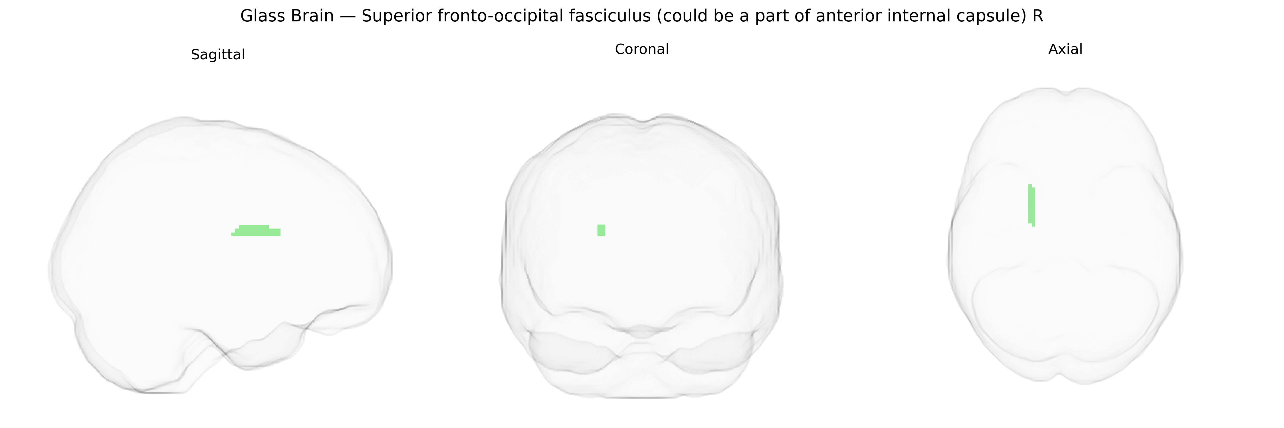
The bilateral Superior fronto-occipital fasciculus (SFOF), as labeled in the JHU ICBM 2mm Atlas and including fibers that may course through the anterior limb of the internal capsule on the right, is a long association white-matter tract that links frontal regions with occipital and parietal cortices. It participates in large-scale cortical networks supporting visuospatial processing, attention, executive functions, and higher-order integration of sensory information by facilitating communication between prefrontal regions and posterior visual association areas. Anatomically, the SFOF runs superior to the corpus callosum and internal capsule, with interindividual variability and some ongoing debate in the literature regarding its exact trajectory and differentiation from neighboring tracts such as the superior longitudinal fasciculus and inferior fronto-occipital fasciculus. There is no direct Wikipedia article for the Superior fronto-occipital fasciculus; a closely related and overlapping structure is the Inferior fronto-occipital fasciculus.
The right superior fronto-occipital fasciculus (SFOF), as defined in the JHU ICBM 2 mm atlas and partly overlapping the anterior limb of the internal capsule, has been implicated in several genetic imaging studies, though often under broader labels such as fronto-occipital or association fibers and internal capsule white matter. Large-scale GWAS of diffusion MRI measures (e.g., fractional anisotropy, mean diffusivity) have identified multiple common variants—many in genes involved in axon guidance, myelination, and neurodevelopment (such as those in or near NRG1, CNTNAP2, and genes in the semaphorin and netrin pathways)—that influence microstructural properties in fronto-occipital and internal capsule tracts, with some loci showing hemispheric or tract-specific effects that likely include the SFOF R. Polygenic risk for schizophrenia, bipolar disorder, and major depressive disorder has been associated with altered integrity of fronto-occipital and internal capsule white matter, and specific risk alleles in genes like ZNF804A, CACNA1C, and DISC1 have been linked to fronto-temporal and fronto-parietal connectivity changes overlapping SFOF pathways. Similarly, GWAS and candidate gene studies of cognitive traits (general intelligence, working memory, processing speed) and neurodevelopmental conditions (autism spectrum disorder, ADHD) report associations between risk variants and microstructural changes in long association tracts connecting frontal and posterior regions, consistent with involvement of the SFOF. In neurodegenerative and vascular disorders, variants in APOE and genes implicated in small-vessel disease (e.g., those highlighted by WMH and stroke GWAS) are associated with white matter hyperintensities and reduced integrity in fronto-occipital and internal capsule regions, again likely encompassing the SFOF R. Overall, genetic associations for this specific labeled tract are typically inferred from broader tract-based or voxelwise analyses rather than SFOF-specific GWAS, but convergent evidence from imaging genetics points to a role for neurodevelopmental, myelination, and vascular risk genes in shaping its structure and its involvement in psychiatric, cognitive, and cerebrovascular phenotypes.
Overview generated by GPT-4o (2026).
Region ID: 43
Hemisphere: bilateral
Atlas: JHU ICBM labels 2mm
Full Quality Version: Download MP4
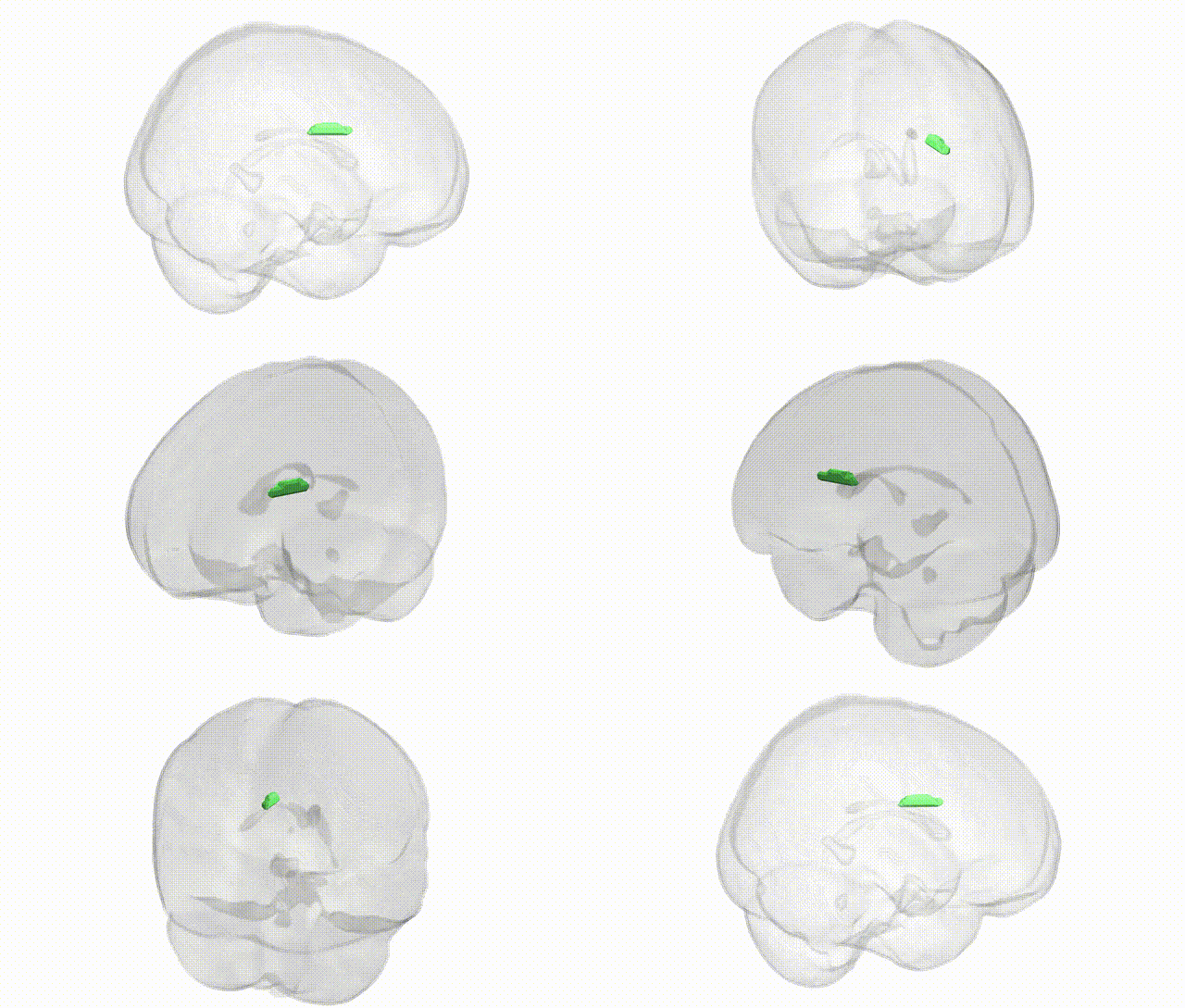
Full Quality Version: Download MP4
Wali Sidiqyar*, Gaurav Rudravaram*, Elyssa M. McMaster, Trent M. Schwartz, Adam M. Saunders, Kurt G. Schilling, Bennett A. Landman "Introducing SPINS: A Shared Public Visualization Library of Neuroanatomical Structures." Medical Imaging with Deep Learning- short paper
This resource is licensed under CC0 1.0 Universal (Public Domain).