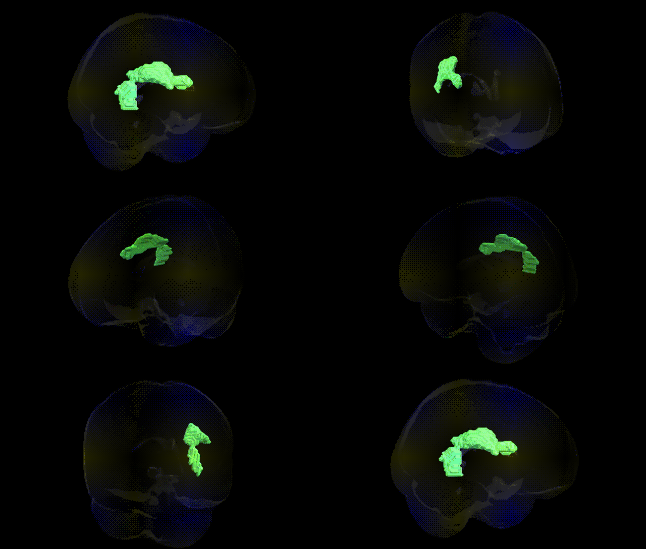
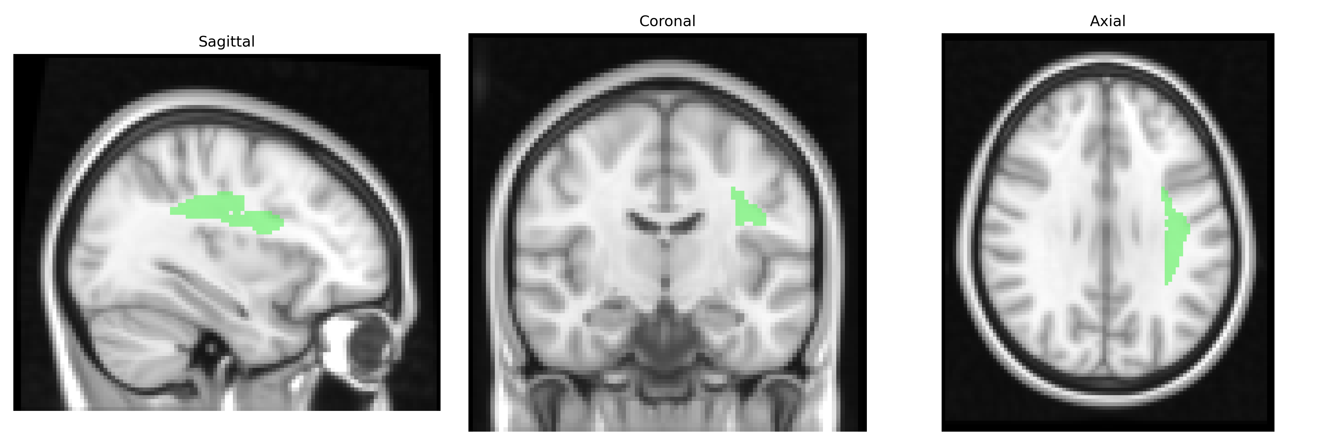
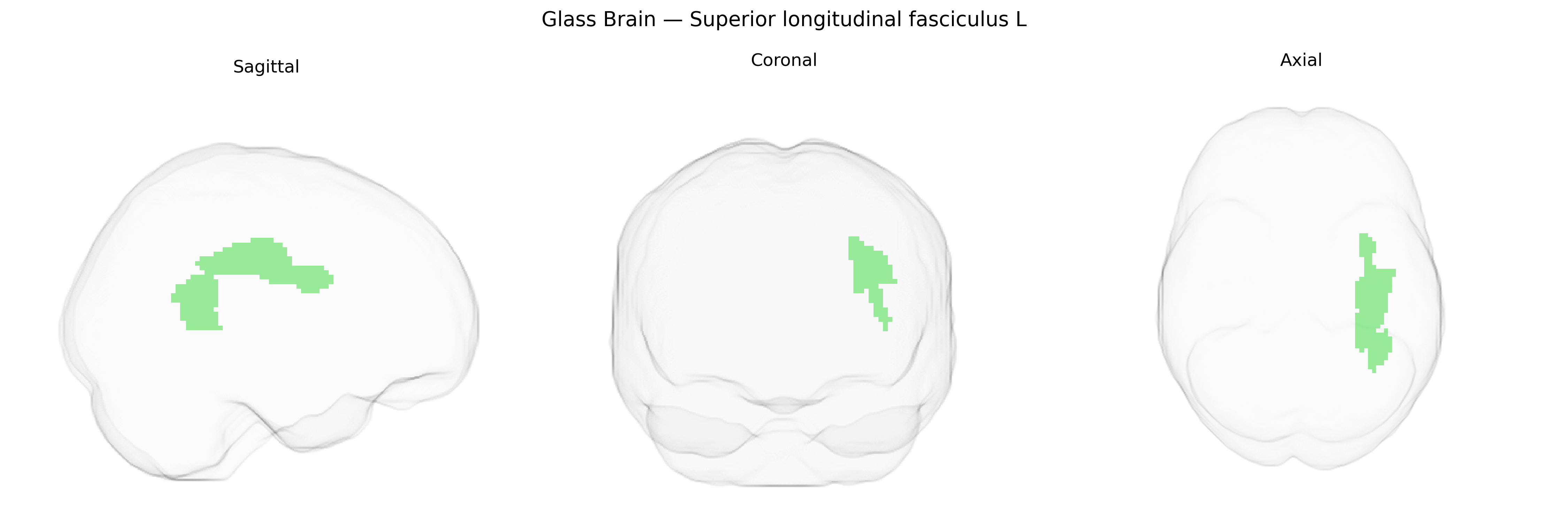
The bilateral Superior longitudinal fasciculus (SLF) is a major associative white matter tract running dorsally within each cerebral hemisphere, connecting frontal lobe regions with parietal, occipital, and temporal cortices. In the JHU ICBM labels 2mm Atlas, the SLF (including the left, or “L,” component) is defined as a long, arching bundle coursing above the insula and lateral ventricles, supporting frontoparietal integration, visuospatial attention, language-related processes, and higher-order cognitive functions. Anatomically, it is often subdivided into distinct segments (e.g., SLF I, II, III and the arcuate component) that link specific cortical territories involved in sensorimotor coordination, spatial awareness, and phonological and semantic aspects of language. There is no direct Wikipedia article for the “Superior longitudinal fasciculus L” label; a related structure is described at Superior longitudinal fasciculus.
The bilateral superior longitudinal fasciculus (SLF) as defined in the JHU ICBM 2 mm atlas has been repeatedly implicated in imaging genetics and GWAS studies of brain structure and connectivity, though loci are generally associated with white matter microstructure (e.g., fractional anisotropy) rather than the SLF label itself. Large-scale diffusion MRI GWAS consortia such as ENIGMA and UK Biobank have identified common variants in genes involved in axon guidance, myelination, and neural development (including but not limited to regions near CNTNAP2, NRG1, MAG, and other oligodendrocyte-related loci) that influence SLF integrity or related frontoparietal tracts. Altered SLF structure has been linked, via polygenic risk or candidate gene approaches, to neurodevelopmental and psychiatric disorders such as schizophrenia, ADHD, autism spectrum disorder, and dyslexia, in which genes affecting synaptic plasticity and white matter development (e.g., CACNA1C, GRM3, FOXP2-related networks) show imaging correlates in SLF connectivity. Additionally, GWAS of cognitive traits—particularly general intelligence, working memory, and language abilities—have reported associations between polygenic scores for cognition and SLF microstructure, with implicated loci enriched in pathways for neurodevelopment, axon growth, and myelin formation. While no single “SLF gene” has been established, convergent evidence indicates that common genetic variation shaping global white matter and frontoparietal circuitry contributes to individual differences in SLF anatomy and to the risk architecture of multiple cognitive and psychiatric phenotypes.
Overview generated by GPT-4o (2026).
Region ID: 42
Hemisphere: bilateral
Atlas: JHU ICBM labels 2mm
Full Quality Version: Download MP4
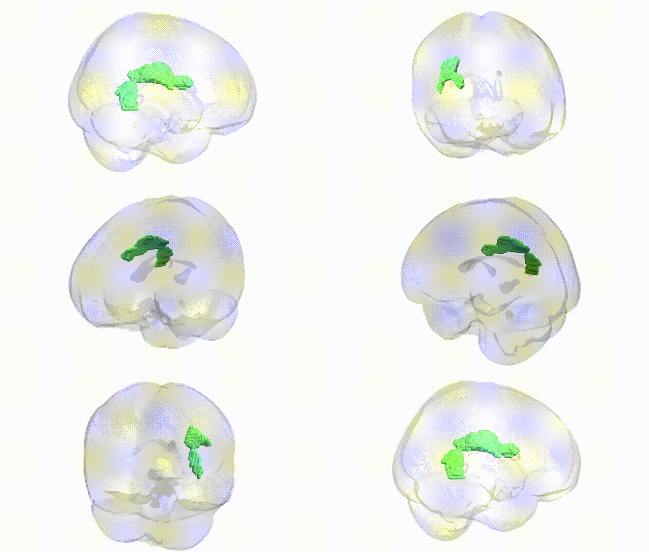
Full Quality Version: Download MP4
Wali Sidiqyar*, Gaurav Rudravaram*, Elyssa M. McMaster, Trent M. Schwartz, Adam M. Saunders, Kurt G. Schilling, Bennett A. Landman "Introducing SPINS: A Shared Public Visualization Library of Neuroanatomical Structures." Medical Imaging with Deep Learning- short paper
This resource is licensed under CC0 1.0 Universal (Public Domain).