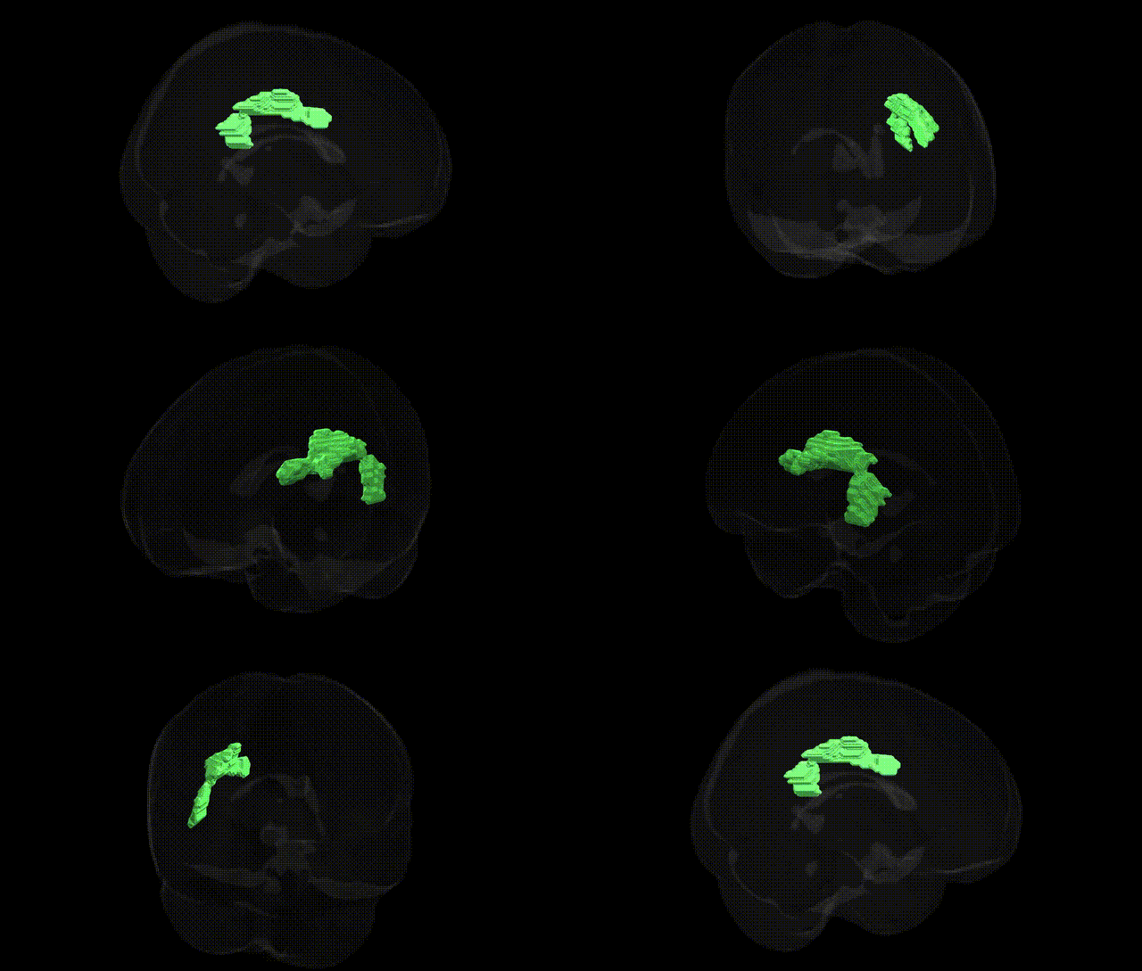
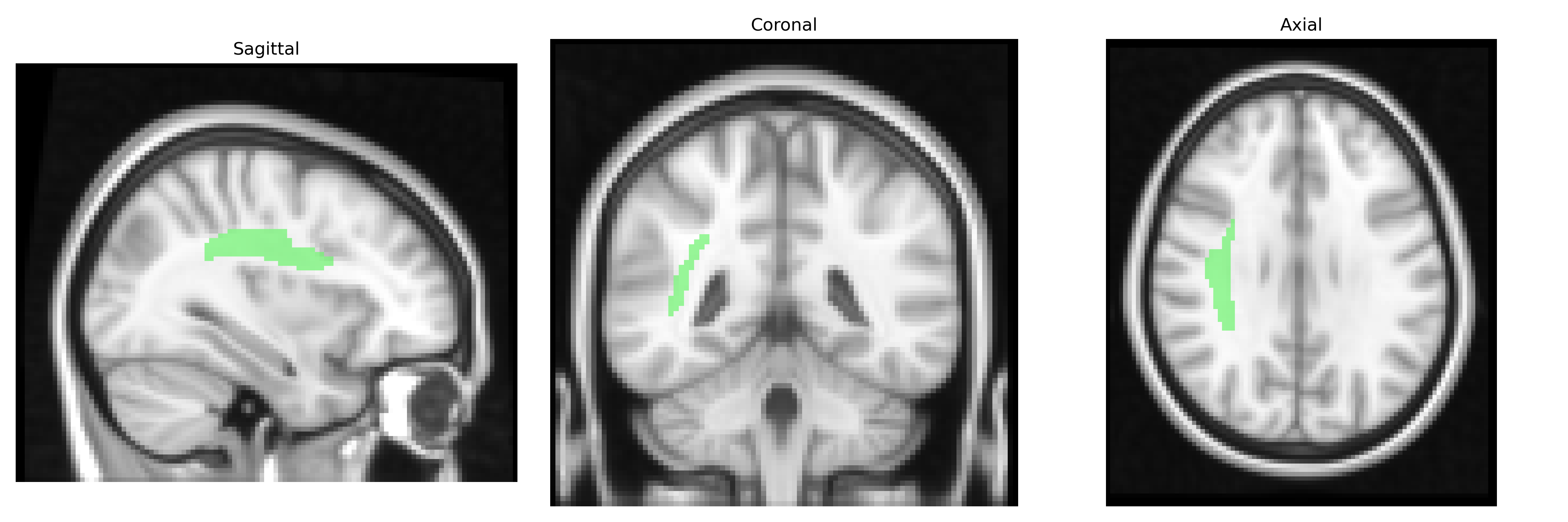
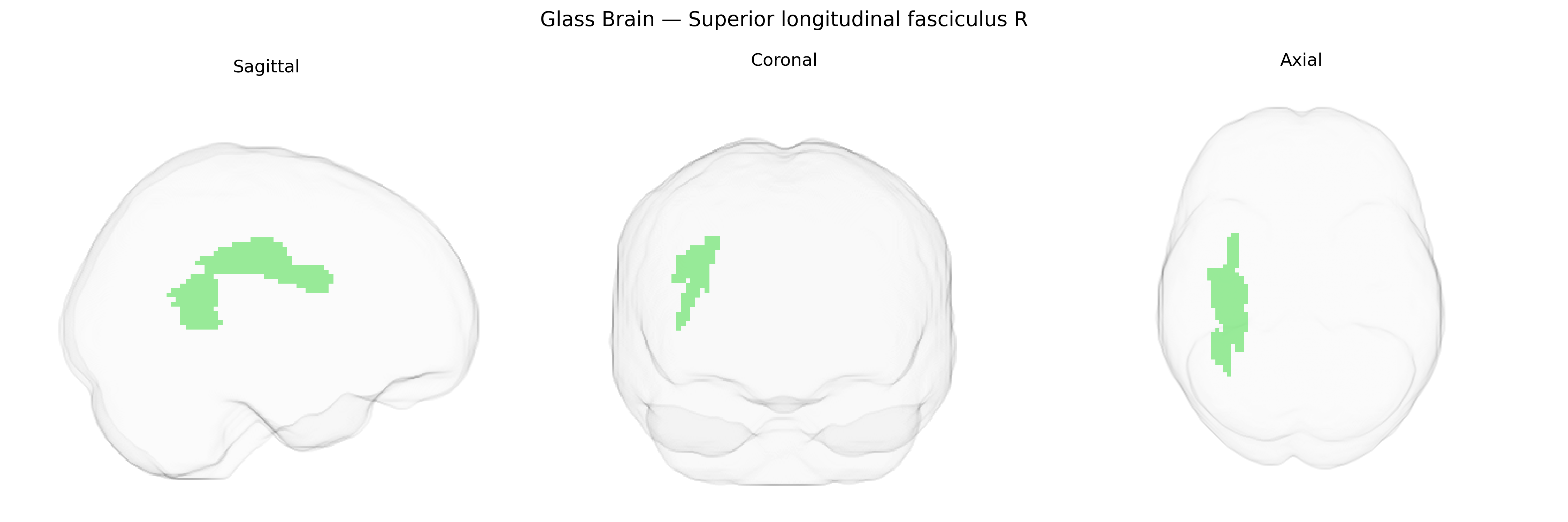
The bilateral Superior longitudinal fasciculus (SLF) is a major associative white matter tract that interconnects frontal, parietal, occipital, and temporal cortical regions, running dorsally and laterally within each hemisphere and playing key roles in attention, visuospatial processing, language, and higher-order cognition. In the JHU ICBM 2mm Atlas, the “Superior longitudinal fasciculus R” refers specifically to the right-hemispheric component of this tract, which links regions such as the inferior parietal lobule and lateral prefrontal cortex, supporting functions including spatial attention and aspects of language and working memory. This fasciculus is composed of several subcomponents (classically SLF I–III and the arcuate fasciculus), which together form an important backbone of long-range cortico-cortical communication. There is no direct Wikipedia article for “Superior longitudinal fasciculus R”; a related entry describing the tract more generally is Superior longitudinal fasciculus.
The bilateral superior longitudinal fasciculus (SLF), including the right SLF as defined in the JHU ICBM labels 2mm atlas, has been repeatedly implicated in imaging genetics and GWAS studies focused on white matter microstructure, cognitive function, and psychiatric risk. Large diffusion MRI GWAS (e.g., UK Biobank–based) have identified multiple common variants influencing fractional anisotropy and other diffusion metrics in SLF tracts, with robust heritability estimates and loci enriched near genes involved in axon guidance, myelination, and cytoskeletal organization (such as those related to neuregulin/ErbB signaling and cell adhesion pathways). Polygenic risk for schizophrenia, bipolar disorder, and major depressive disorder has been associated with altered SLF integrity, particularly in fronto-parietal segments, and rare copy number variants (e.g., 22q11.2 deletion) also show convergent effects on SLF structure. Variants in genes including CNTNAP2, DISC1, and NRG1, as well as FOXP2-related networks, have been linked to language and reading abilities along with white matter changes in SLF, supporting its role in verbal working memory, phonological processing, and broader cognitive performance. ADHD and autism spectrum disorder risk alleles have likewise been associated with SLF differences in some imaging genetics studies, consistent with the tract’s involvement in attention, executive function, and social cognition, though specific genome-wide significant loci tied uniquely and exclusively to the JHU-defined right SLF remain limited and are usually reported at the level of broader fronto-parietal white matter tracts rather than this single atlas-defined region.
Overview generated by GPT-4o (2026).
Region ID: 41
Hemisphere: bilateral
Atlas: JHU ICBM labels 2mm
Full Quality Version: Download MP4
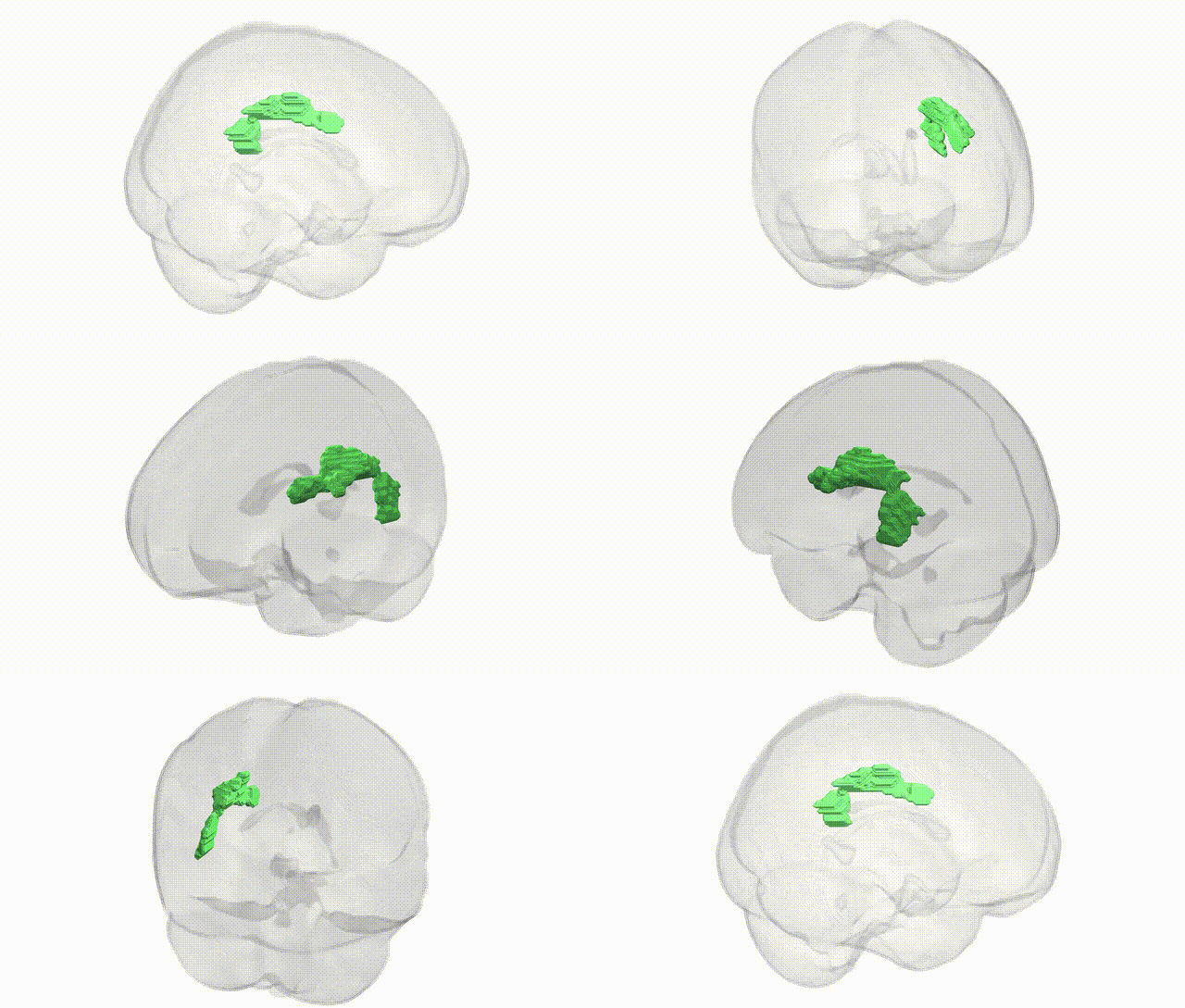
Full Quality Version: Download MP4
Wali Sidiqyar*, Gaurav Rudravaram*, Elyssa M. McMaster, Trent M. Schwartz, Adam M. Saunders, Kurt G. Schilling, Bennett A. Landman "Introducing SPINS: A Shared Public Visualization Library of Neuroanatomical Structures." Medical Imaging with Deep Learning- short paper
This resource is licensed under CC0 1.0 Universal (Public Domain).