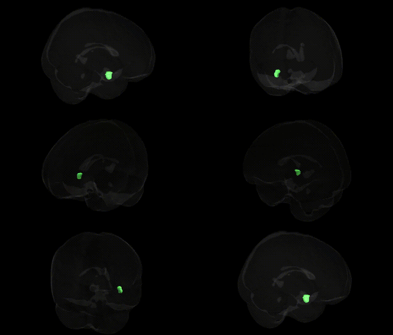
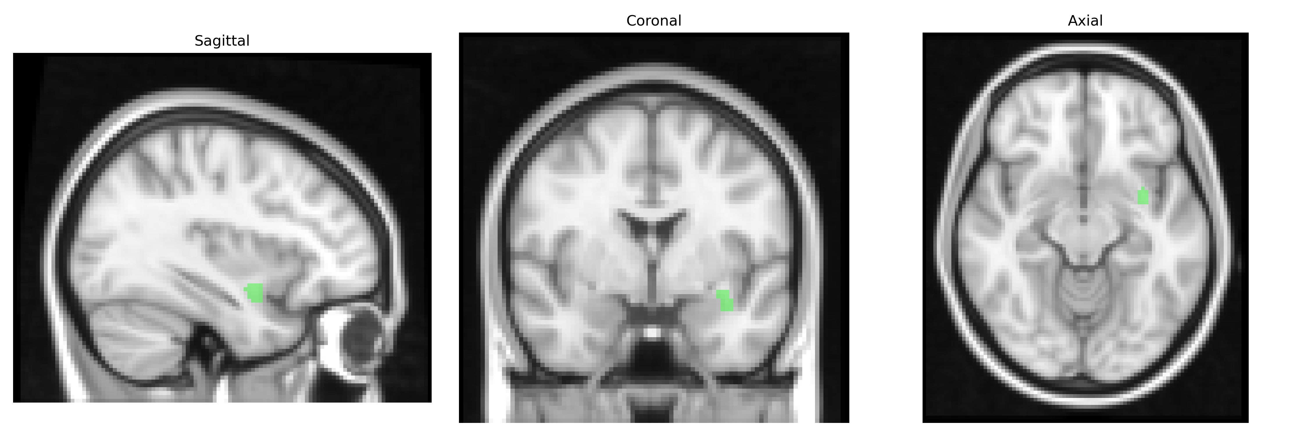
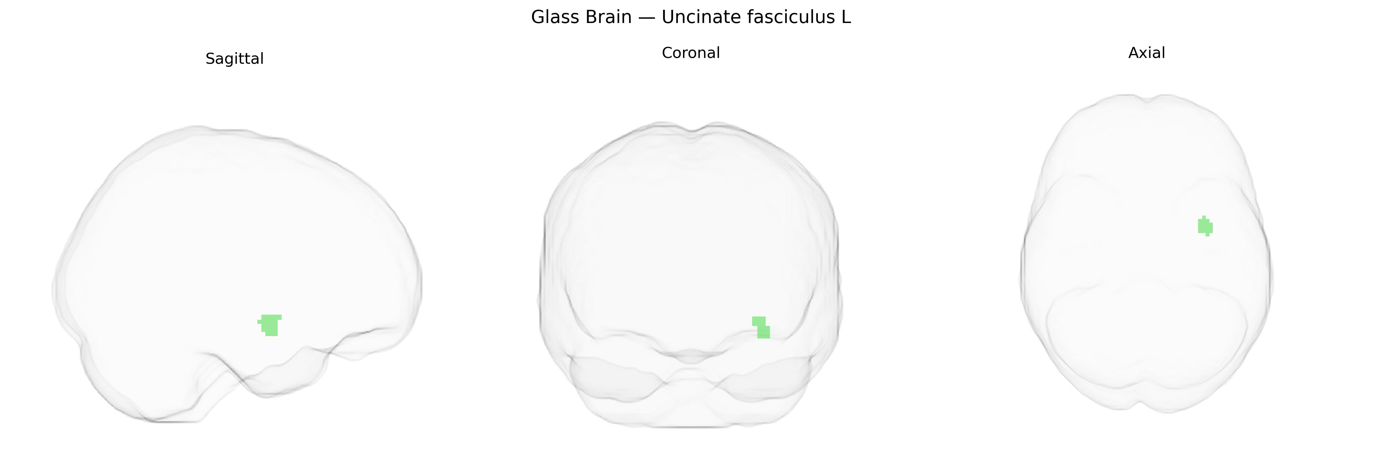
The bilateral Uncinate fasciculus L, as defined in the JHU ICBM labels 2mm Atlas, is a left-lateralized major associative white matter tract that connects anterior temporal lobe structures, including the temporal pole and amygdalo-hippocampal regions, with the orbitofrontal and ventromedial prefrontal cortices via a characteristically hook-shaped (uncinate) trajectory around the Sylvian fissure. Composed of heavily myelinated fibers running within the deep white matter of the frontal and temporal lobes, it is implicated in higher-order cognitive and affective functions such as episodic memory, semantic processing, emotional regulation, and social cognition, and is frequently studied in diffusion tensor imaging and tractography research for its role in neurodevelopmental, neuropsychiatric, and neurodegenerative disorders. Uncinate fasciculus
The bilateral uncinate fasciculus, a frontotemporal white-matter tract labeled in the JHU ICBM 2 mm atlas, has been implicated in multiple genetic and GWAS findings primarily through imaging-genetics and disorder-based studies rather than direct tract-specific GWAS hits. Variants in genes influencing myelination and axonal integrity—such as those in the NTRK1/3, CNTNAP2, and neuregulin pathways—have been associated with microstructural measures (e.g., fractional anisotropy) in frontotemporal tracts including the uncinate fasciculus, although many findings remain exploratory. Polygenic risk scores for schizophrenia, bipolar disorder, and major depressive disorder have been linked to altered uncinate fasciculus integrity, and risk loci in genes like ZNF804A and CACNA1C have shown associations with white-matter abnormalities that prominently involve fronto-limbic pathways. In neurodevelopmental conditions such as autism spectrum disorder and ADHD, GWAS and candidate gene studies (e.g., involving CNTNAP2, DISC1, and FOXP2-related networks) have been connected to disrupted frontotemporal connectivity including the uncinate fasciculus. Alzheimer’s disease and frontotemporal dementia risk genes (such as APOE and MAPT) have also been tied to degeneration of the uncinate fasciculus in imaging-genetic studies. Overall, genetic influences on the uncinate fasciculus appear highly polygenic and shared across psychiatric and neurodegenerative disorders, with GWAS signals typically identified at the level of global or regional white-matter metrics rather than as tract-specific associations confined to the bilateral uncinate fasciculus label in the JHU ICBM atlas.
Overview generated by GPT-4o (2026).
Region ID: 46
Hemisphere: bilateral
Atlas: JHU ICBM labels 2mm
Full Quality Version: Download MP4
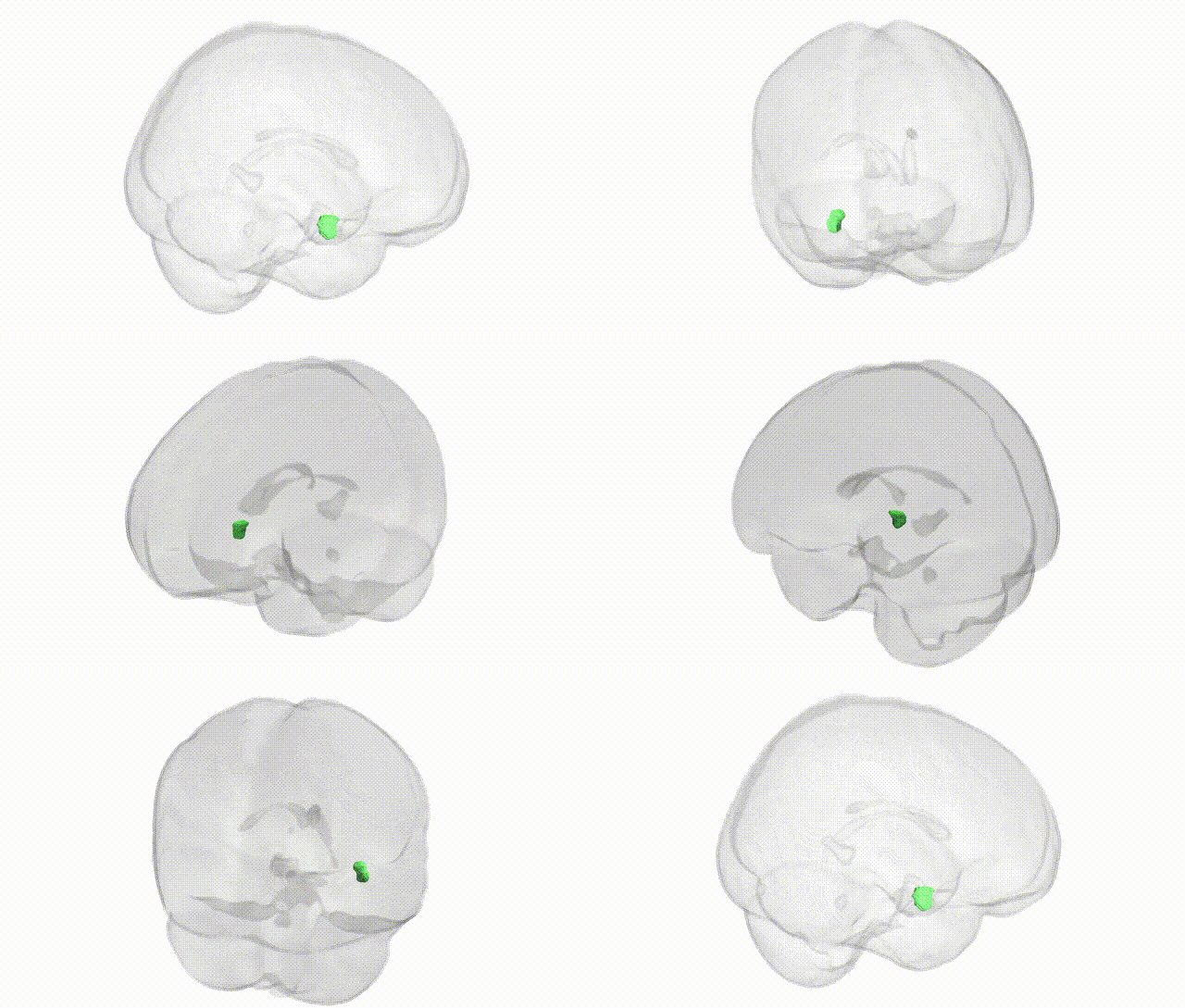
Full Quality Version: Download MP4
Wali Sidiqyar*, Gaurav Rudravaram*, Elyssa M. McMaster, Trent M. Schwartz, Adam M. Saunders, Kurt G. Schilling, Bennett A. Landman "Introducing SPINS: A Shared Public Visualization Library of Neuroanatomical Structures." Medical Imaging with Deep Learning- short paper
This resource is licensed under CC0 1.0 Universal (Public Domain).