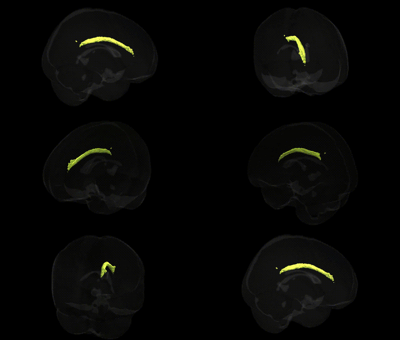
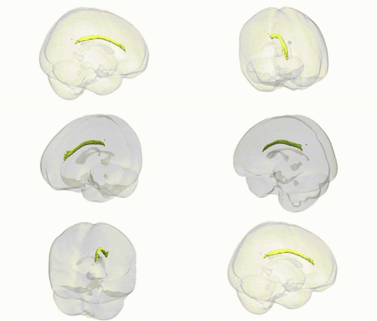
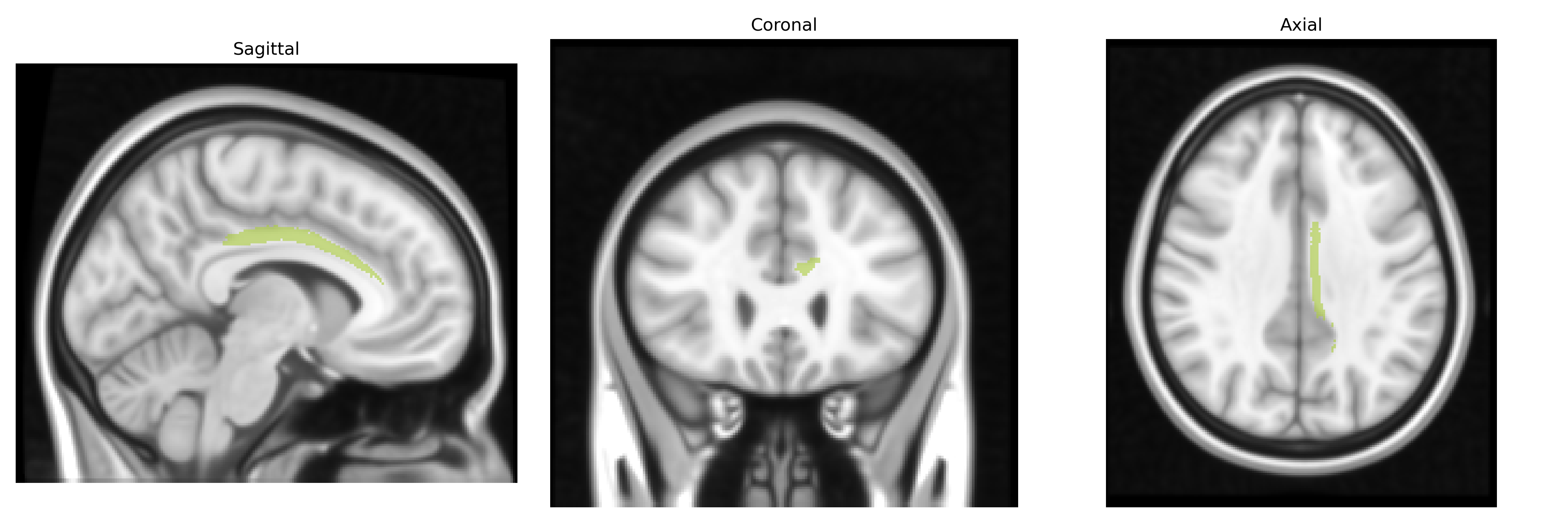
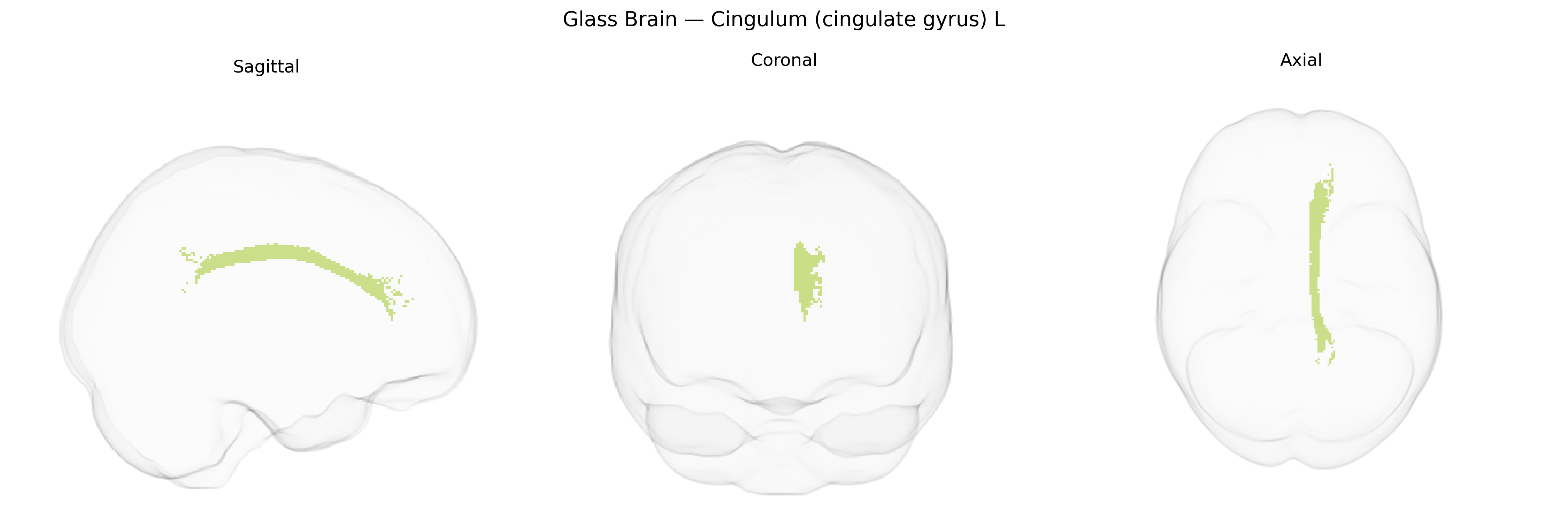
The bilateral Cingulum (cingulate gyrus) L, as defined in the JHU ICBM tracts maxprob thr25 1 mm atlas, refers to the left-lateralized segment of the cingulum white matter tract coursing within and beneath the cingulate gyrus, arching around the corpus callosum and extending toward medial temporal structures. This tract forms a major component of the limbic system’s structural connectivity, linking medial frontal, cingulate, parietal, and parahippocampal regions, and is implicated in the integration of cognitive control, attention, emotional processing, and memory-related functions. The cingulum’s fibers support bidirectional communication between the anterior cingulate cortex, posterior cingulate cortex/precuneus, and medial temporal lobe, contributing to large-scale networks such as the default mode network and playing a role in adaptive behavior, affect regulation, and the coordination of internally directed mentation.
The bilateral cingulum (cingulate gyrus) tract, as defined in the JHU ICBM tracts maxprob thr25 1mm atlas, has been repeatedly implicated in genetic studies of brain structure and neuropsychiatric risk, with GWAS and imaging‑genetics work showing heritability of its microstructural properties (e.g., fractional anisotropy, mean diffusivity) and polygenic influences from loci associated with neurodevelopment and synaptic function. Large consortia such as ENIGMA and UK Biobank have reported associations between common variants in genes including those related to axonal guidance (e.g., ROBO/SLIT pathways), myelination (e.g., genes near MAG, MBP regions), and neurodevelopmental regulation with cingulum diffusion metrics, often in concert with global white‑matter genetic factors. The cingulum’s connectivity with limbic and frontal networks has made it a focus of imaging‑genetics studies in major depressive disorder, anxiety, schizophrenia, bipolar disorder, and ADHD, where polygenic risk scores for these conditions correlate with altered cingulum microstructure or volume, suggesting that shared genetic architectures influence both tract integrity and vulnerability to psychiatric illness. In Alzheimer’s disease and other dementias, variants in APOE and genes involved in amyloid and tau pathways have been linked to degeneration and reduced integrity of cingulum fibers, particularly in posterior sections, reflecting genetic contributions to white‑matter changes in neurodegeneration. Additional genetic associations involve traits such as neuroticism, cognitive performance, and emotional regulation, where GWAS-derived polygenic scores correlate with cingulum properties, supporting the view that this tract is a genetically modulated hub connecting cognition, emotion, and psychopathology.
Overview generated by GPT-4o (2026).
Region ID: 5
Hemisphere: bilateral
Atlas: JHU ICBM tracts maxprob thr25 1mm
Full Quality Version: Download MP4
Full Quality Version: Download MP4
Wali Sidiqyar*, Gaurav Rudravaram*, Elyssa M. McMaster, Trent M. Schwartz, Adam M. Saunders, Kurt G. Schilling, Bennett A. Landman "Introducing SPINS: A Shared Public Visualization Library of Neuroanatomical Structures." Medical Imaging with Deep Learning- short paper
This resource is licensed under CC0 1.0 Universal (Public Domain).