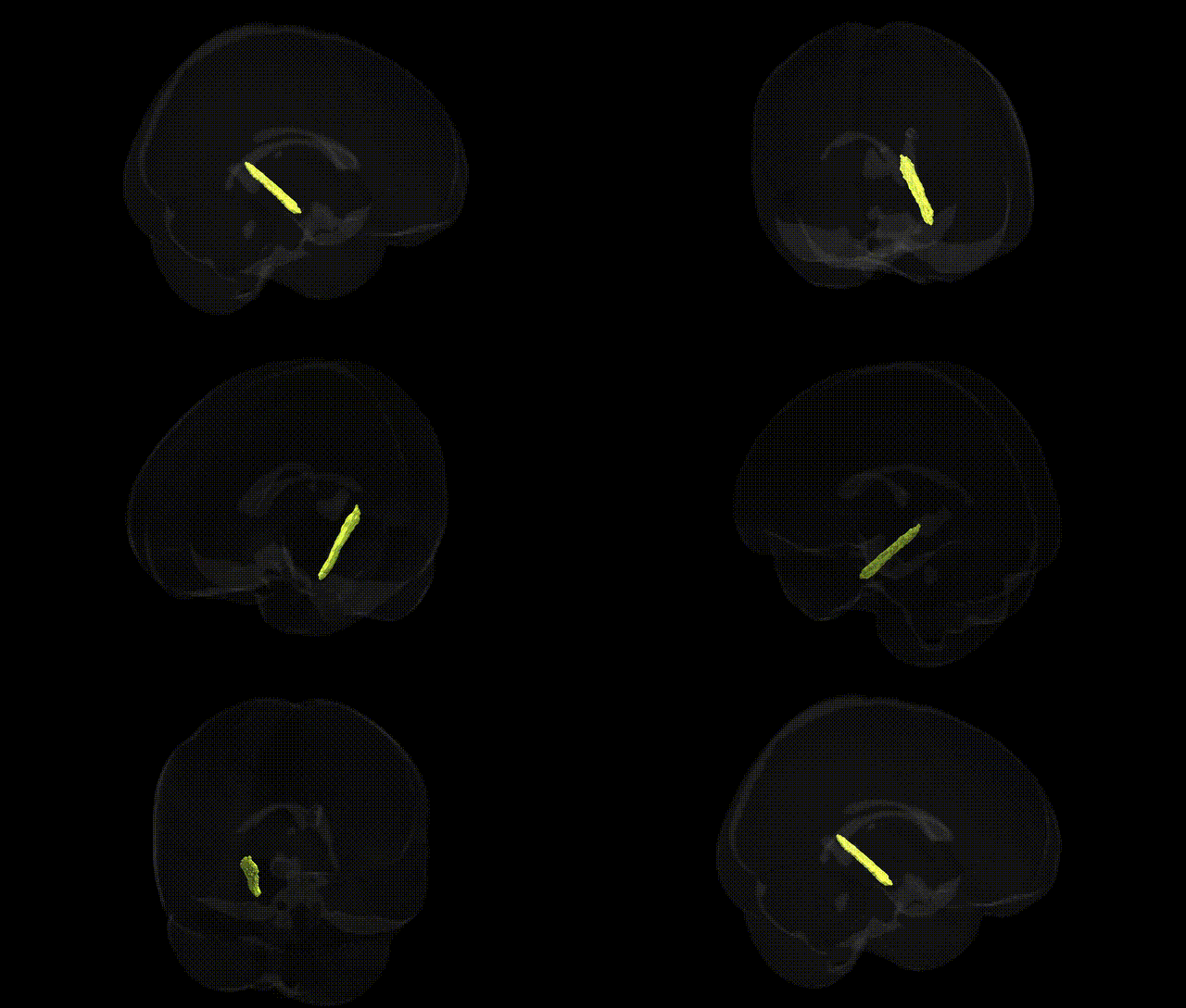
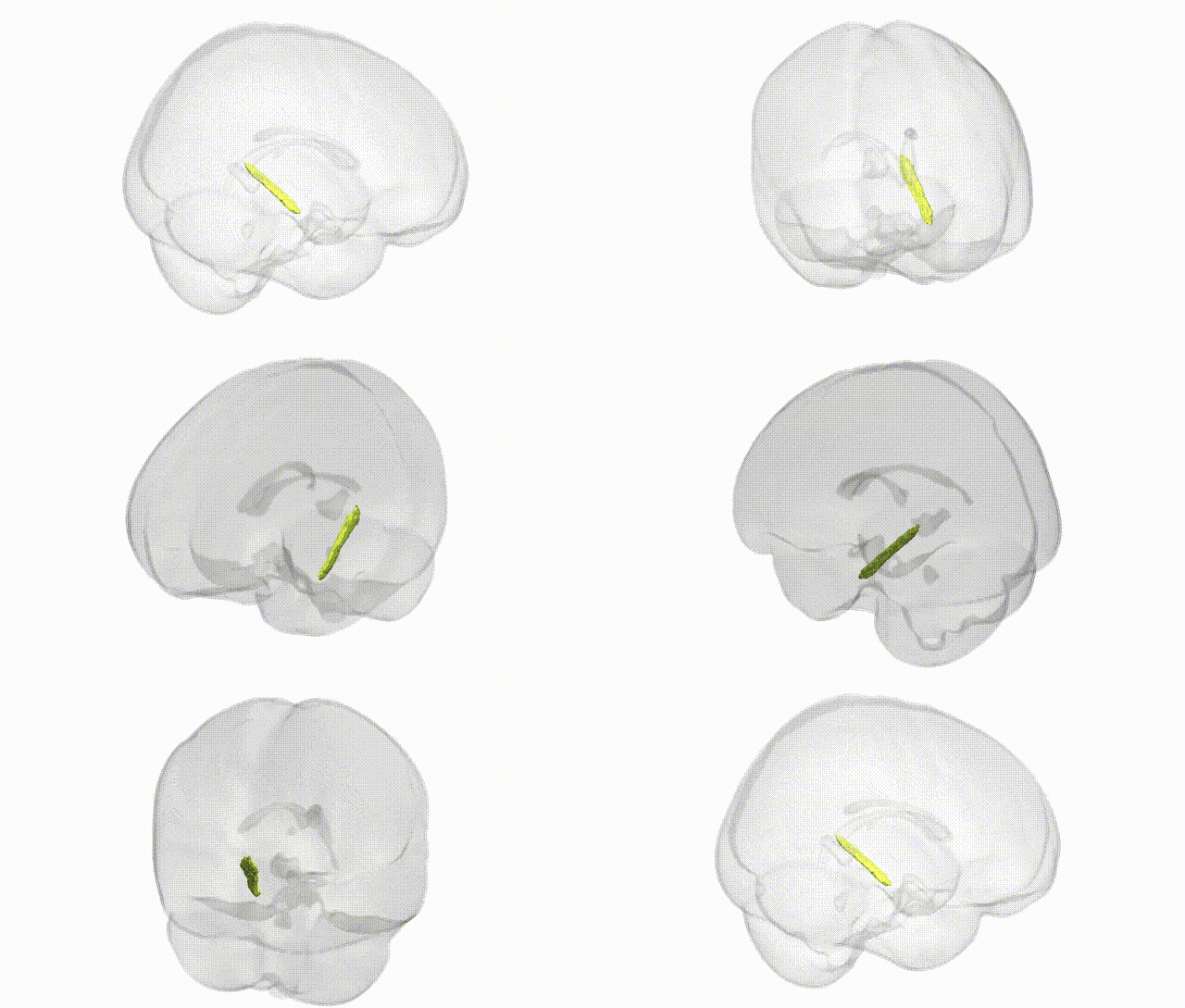
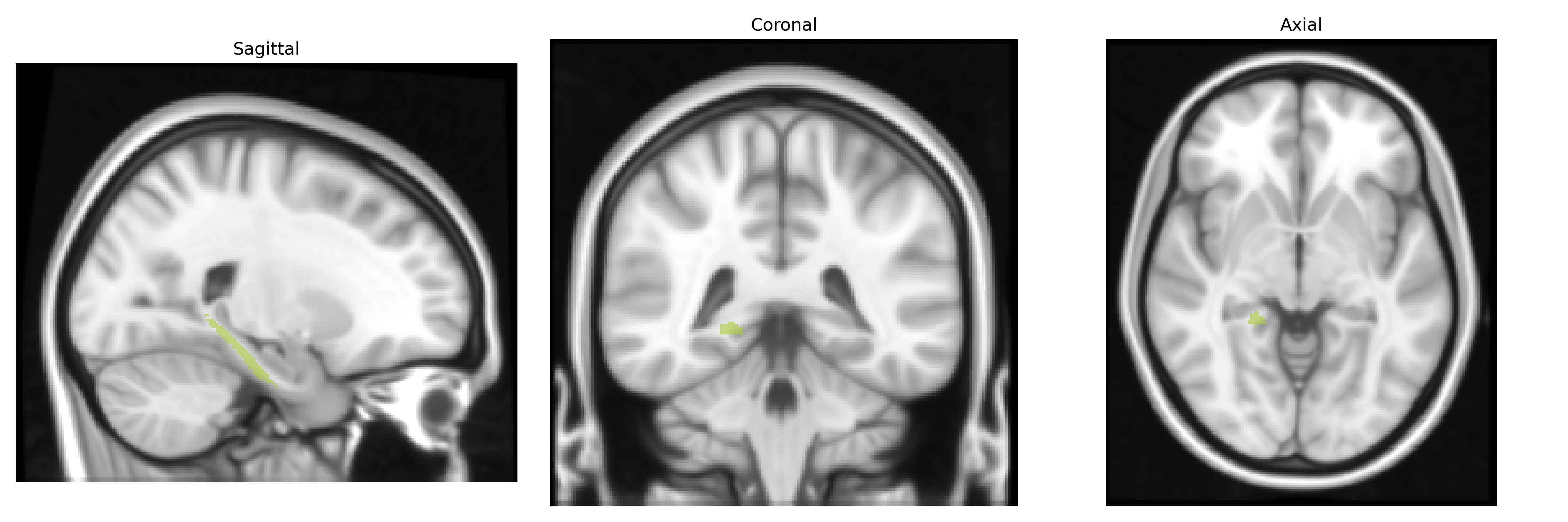
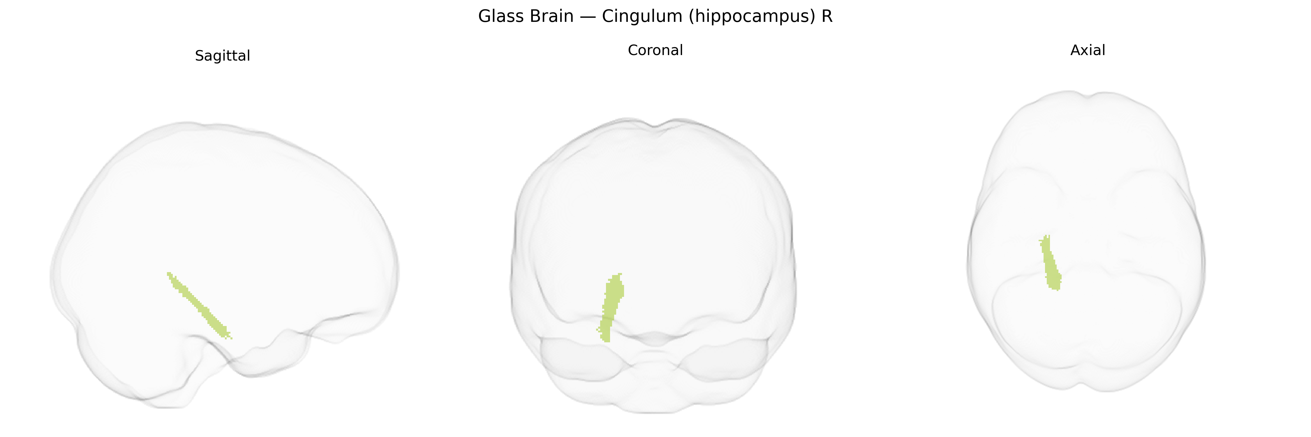
The bilateral Cingulum (hippocampus) R, as defined in the JHU ICBM tracts maxprob thr25 1mm Atlas, refers to the hippocampal segment of the cingulum bundle coursing within the parahippocampal region and connecting medial temporal lobe structures, including the hippocampus, with medial parietal and frontal association cortices. This white matter tract participates in limbic circuitry, supporting episodic memory, spatial navigation, and integration of emotional and contextual information by linking the hippocampal formation with the cingulate gyrus and other components of the limbic system. Its fibers run longitudinally along the parahippocampal gyrus and contribute to large-scale networks such as the default mode network, making it critical for coherent communication between memory-related temporal structures and higher-order cortical regions. Cingulum bundle
Genetic associations with microstructure of the bilateral cingulum bundle adjacent to the hippocampus (as defined in the JHU ICBM tracts atlas) have been identified primarily through diffusion MRI GWAS, which implicate common variants influencing fractional anisotropy and other white matter metrics in this tract; notable findings include loci near or within genes involved in neurodevelopment, myelination, and axon guidance (such as those in pathways overlapping with NTRK2/BDNF signaling and cell-adhesion molecules), although specific single-gene effects are typically small and polygenic. Large-scale imaging-genetics consortia (for example ENIGMA and UK Biobank–based studies) report that cingulum-hippocampal diffusion measures exhibit significant SNP-based heritability and share genetic correlations with cognitive performance, educational attainment, and general brain structural integrity. In case–control and polygenic score analyses, altered cingulum (hippocampus) microstructure has been linked to risk for major depressive disorder, schizophrenia, bipolar disorder, and post-traumatic stress disorder, with some overlap between GWAS hits for these disorders and variants influencing white matter metrics in this tract, suggesting a shared polygenic architecture rather than single-region-specific “risk genes.” Additionally, neurodegenerative and aging-related genetic risk—such as APOE ε4 and polygenic scores for Alzheimer’s disease—has been associated with reduced integrity of the hippocampal cingulum, consistent with its role in memory networks and vulnerability to age-related and pathological processes.
Overview generated by GPT-4o (2026).
Region ID: 8
Hemisphere: bilateral
Atlas: JHU ICBM tracts maxprob thr25 1mm
Full Quality Version: Download MP4
Full Quality Version: Download MP4
Wali Sidiqyar*, Gaurav Rudravaram*, Elyssa M. McMaster, Trent M. Schwartz, Adam M. Saunders, Kurt G. Schilling, Bennett A. Landman "Introducing SPINS: A Shared Public Visualization Library of Neuroanatomical Structures." Medical Imaging with Deep Learning- short paper
This resource is licensed under CC0 1.0 Universal (Public Domain).