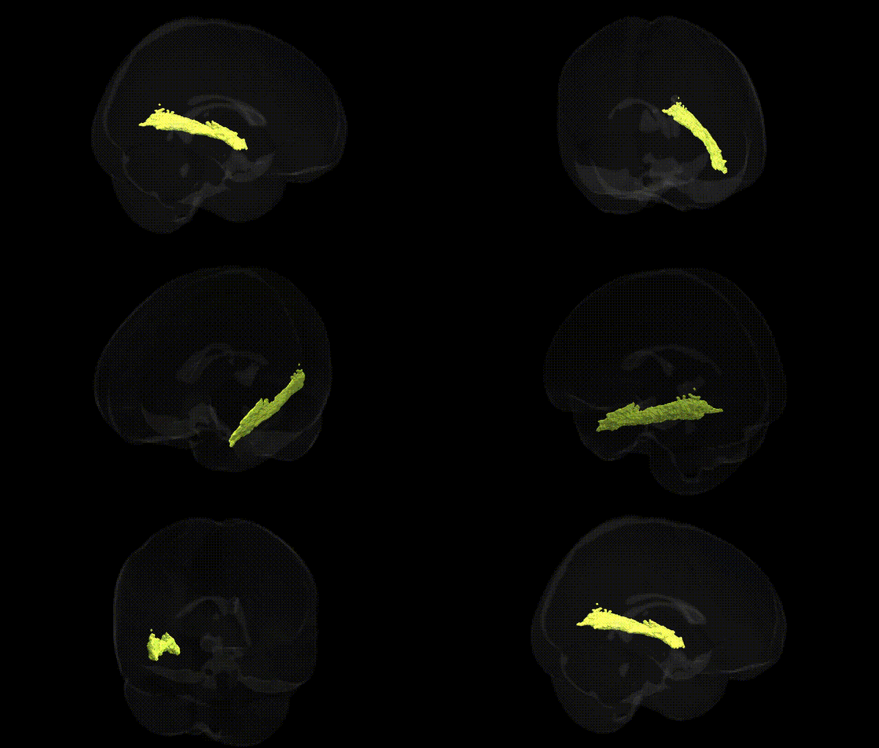
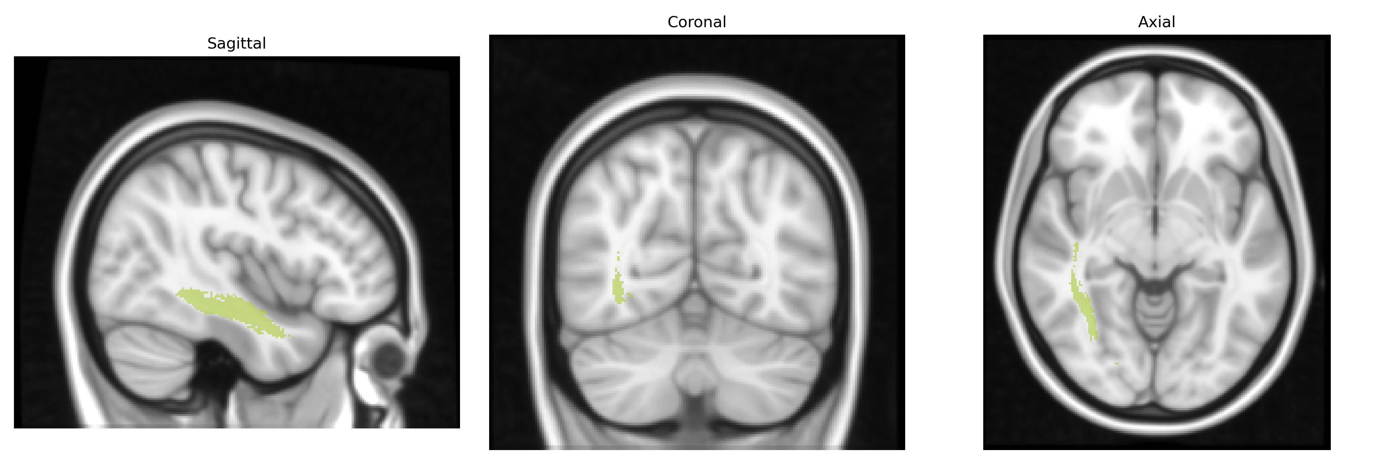
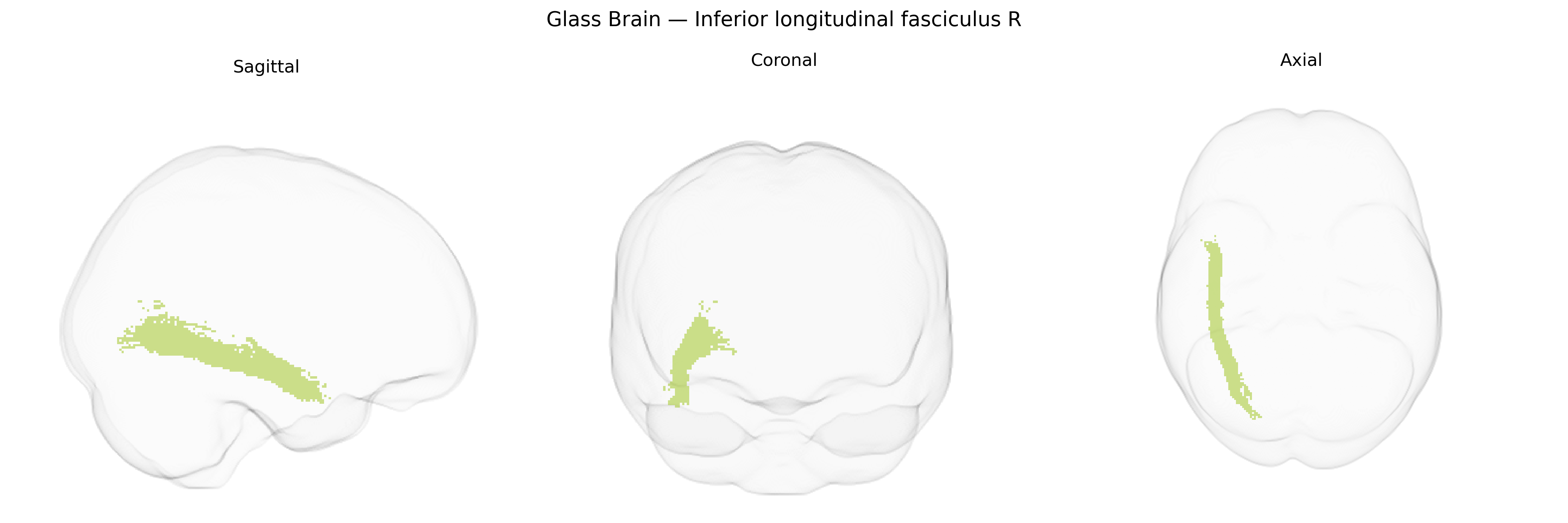
The bilateral Inferior Longitudinal Fasciculus (ILF) is a major associative white matter tract that extends along the ventral temporal pathway, connecting occipital visual areas to anterior and medial temporal regions, including portions of the fusiform gyrus and temporal pole. It runs longitudinally beneath the lateral ventricles and the cortical surface of the temporal lobe, playing a critical role in visual object recognition, face processing, reading, and the integration of visual information with semantic and emotional processing. In the JHU ICBM tracts maxprob thr25 1mm atlas, this tract is delineated probabilistically in both hemispheres, reflecting its bilateral organization and facilitating studies of structure–function relationships in cognition and perception. There is no dedicated Wikipedia article specifically for the ILF; a related and encompassing structure is the white matter of the brain: White matter.
The bilateral inferior longitudinal fasciculus (ILF), a major occipito-temporal association tract represented in the JHU ICBM tracts maxprob thr25 1 mm Atlas (including the right ILF segment), has been implicated in multiple imaging genetics and GWAS-based endophenotype studies, though few findings target this tract in isolation. Diffusion MRI GWAS of white matter microstructure (e.g., using fractional anisotropy and mean diffusivity) have identified common variants in genes related to axon guidance, myelination, and neural development—such as those in the netrin, semaphorin, and neuregulin signaling pathways—that modulate association tracts including the ILF. ILF microstructure has been genetically associated, at the polygenic or locus level, with risk for schizophrenia, bipolar disorder, autism spectrum disorder, and major depressive disorder, where risk alleles linked to synaptic plasticity and neurodevelopment (e.g., in CACNA1C, NRG1, and other neurodevelopmental genes) show correlates in reduced integrity or atypical lateralization of temporal–occipital tracts. Imaging-genetics work has also connected ILF characteristics to cognitive and behavioral traits such as reading ability, language and semantic processing, face and object recognition, and broader intelligence factors, often through polygenic scores for educational attainment and cognitive performance that predict ILF microstructural variation. Additionally, ILF integrity, including in the right hemisphere, has been reported in genetically informed studies of neurodevelopmental disorders (dyslexia, ADHD) and neurodegeneration (Alzheimer’s disease risk variants like APOE), suggesting that common genetic variation influencing cortical development, myelination, and large-scale network connectivity can exert measurable effects on this tract, although the field still lacks large, ILF-specific GWAS with robust, tract-focused loci.
Overview generated by GPT-4o (2026).
Region ID: 14
Hemisphere: bilateral
Atlas: JHU ICBM tracts maxprob thr25 1mm
Full Quality Version: Download MP4
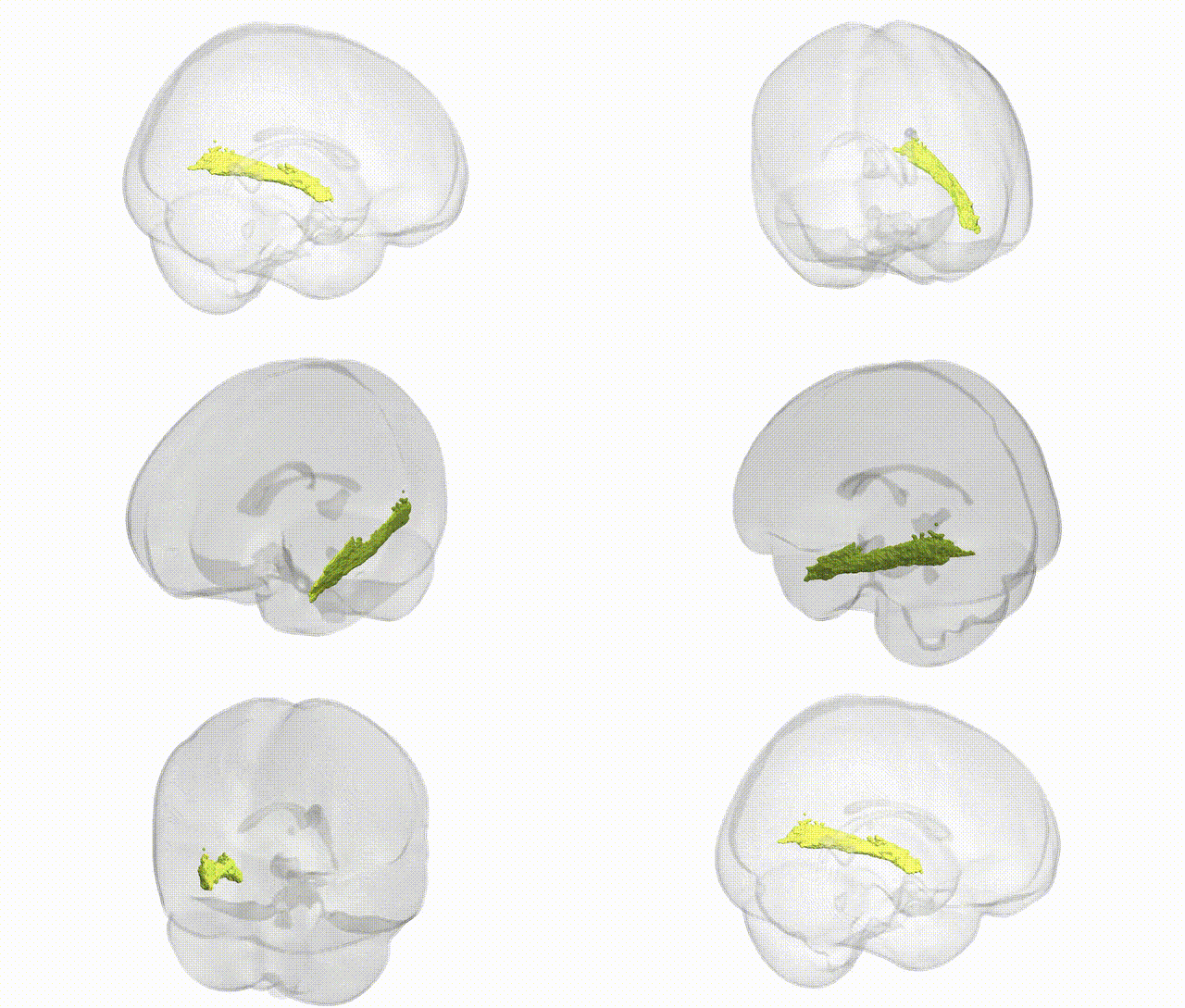
Full Quality Version: Download MP4
Wali Sidiqyar*, Gaurav Rudravaram*, Elyssa M. McMaster, Trent M. Schwartz, Adam M. Saunders, Kurt G. Schilling, Bennett A. Landman "Introducing SPINS: A Shared Public Visualization Library of Neuroanatomical Structures." Medical Imaging with Deep Learning- short paper
This resource is licensed under CC0 1.0 Universal (Public Domain).