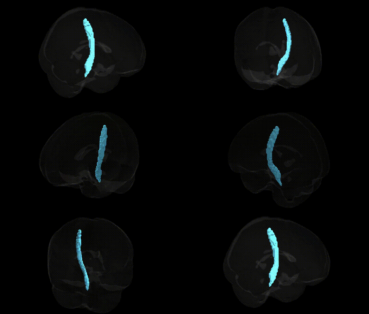
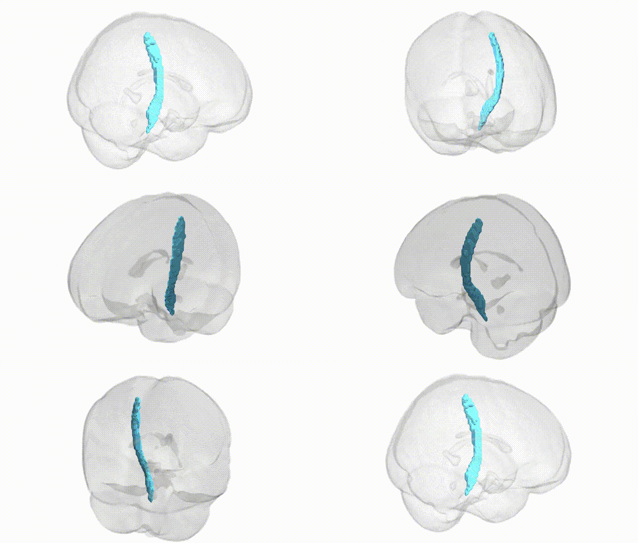
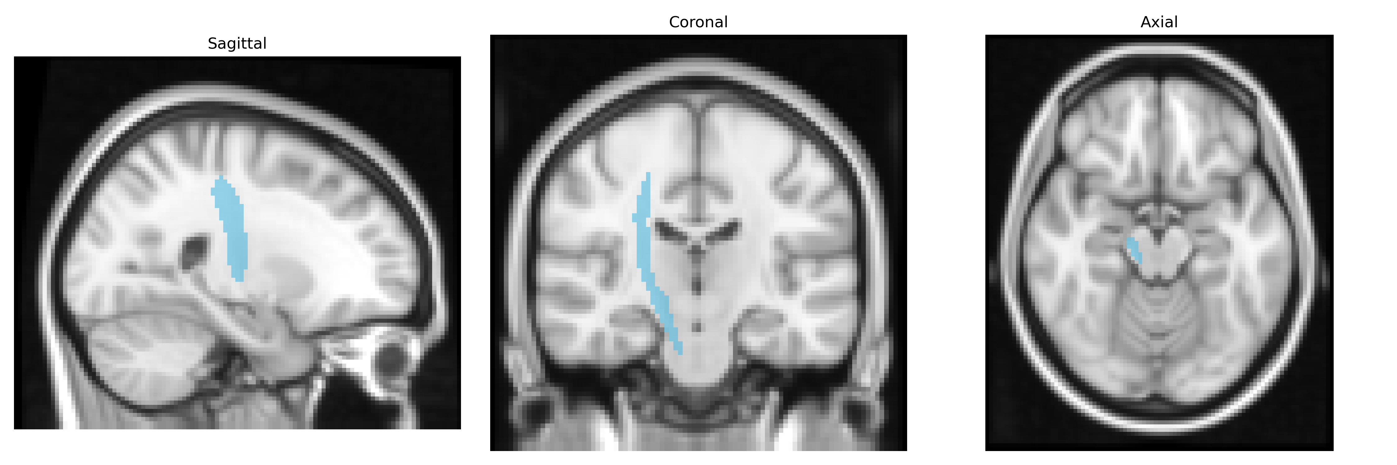
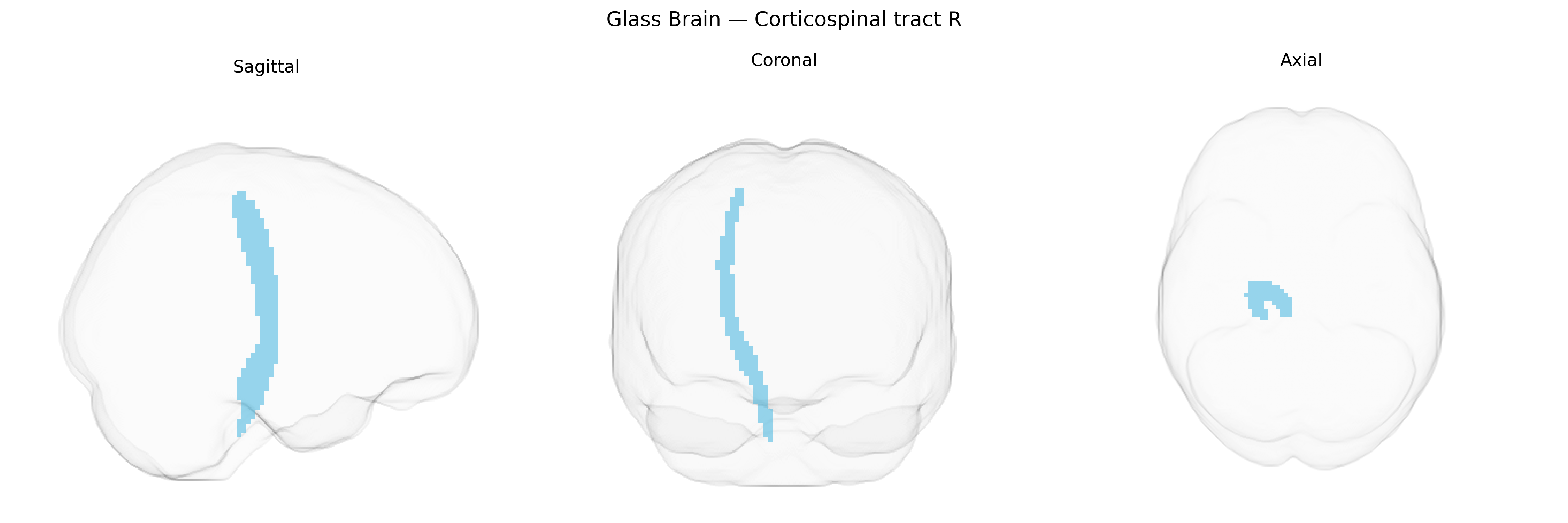
The bilateral corticospinal tract (right) is a major descending white matter pathway that originates primarily from pyramidal neurons in the primary motor cortex and additional motor-related areas of the frontal lobe, then travels through the corona radiata and posterior limb of the internal capsule, continuing caudally through the cerebral peduncles, basis pontis, and medullary pyramids before most fibers decussate in the caudal medulla to innervate contralateral spinal motor circuits. This tract is the principal conduit for voluntary, fine, and fractionated movement control of the limbs and trunk, transmitting motor commands to alpha and gamma motor neurons and interneurons in the spinal cord ventral horn. Damage to the corticospinal tract can cause characteristic upper motor neuron signs, including weakness, spasticity, hyperreflexia, and loss of fine motor control. There is no direct link for the “corticospinal tract” as a standalone article; a closely related and encompassing structure is described under Pyramidal tracts.
Genetic associations involving the bilateral corticospinal tract (right) as defined in the JHU ICBM tracts maxprob thr25 2mm atlas arise mainly from imaging-genetics and GWAS of white matter microstructure rather than tract-specific gene mapping. Heritability studies using diffusion tensor imaging indicate substantial genetic contribution to corticospinal tract integrity (e.g., fractional anisotropy and mean diffusivity), with polygenic influences distributed across many loci. GWAS of global and regional white matter measures have implicated genes involved in axon guidance, myelination, and cytoskeletal organization—such as CNTN4, ROBO1/2, and myelin-related genes like MAG and MBP—as broadly relevant to long-range motor pathways that include the corticospinal tract, though associations are generally not specific to the right tract alone. Variants in genes linked to neurodevelopmental and motor disorders (e.g., ALS-associated genes like C9orf72, SOD1, and FUS; cerebral palsy susceptibility loci; and genes impacting motor cortex development) have been associated with structural or functional changes in motor pathways encompassing the corticospinal tract in case–control imaging-genetics studies. Additionally, polygenic risk for neuropsychiatric and neurodevelopmental conditions (such as schizophrenia, ADHD, and autism spectrum disorder) has been correlated with alterations in major white matter tracts, including the corticospinal tract, suggesting pleiotropic influences of common variants on motor and higher-order networks, though current evidence points to diffuse, non-tract-specific genetic architecture rather than strong, unique GWAS hits confined to the right corticospinal tract region.
Overview generated by GPT-4o (2026).
Region ID: 4
Hemisphere: bilateral
Atlas: JHU ICBM tracts maxprob thr25 2mm
Full Quality Version: Download MP4
Full Quality Version: Download MP4
Wali Sidiqyar*, Gaurav Rudravaram*, Elyssa M. McMaster, Trent M. Schwartz, Adam M. Saunders, Kurt G. Schilling, Bennett A. Landman "Introducing SPINS: A Shared Public Visualization Library of Neuroanatomical Structures." Medical Imaging with Deep Learning- short paper
This resource is licensed under CC0 1.0 Universal (Public Domain).