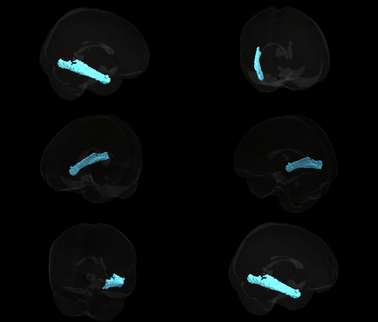
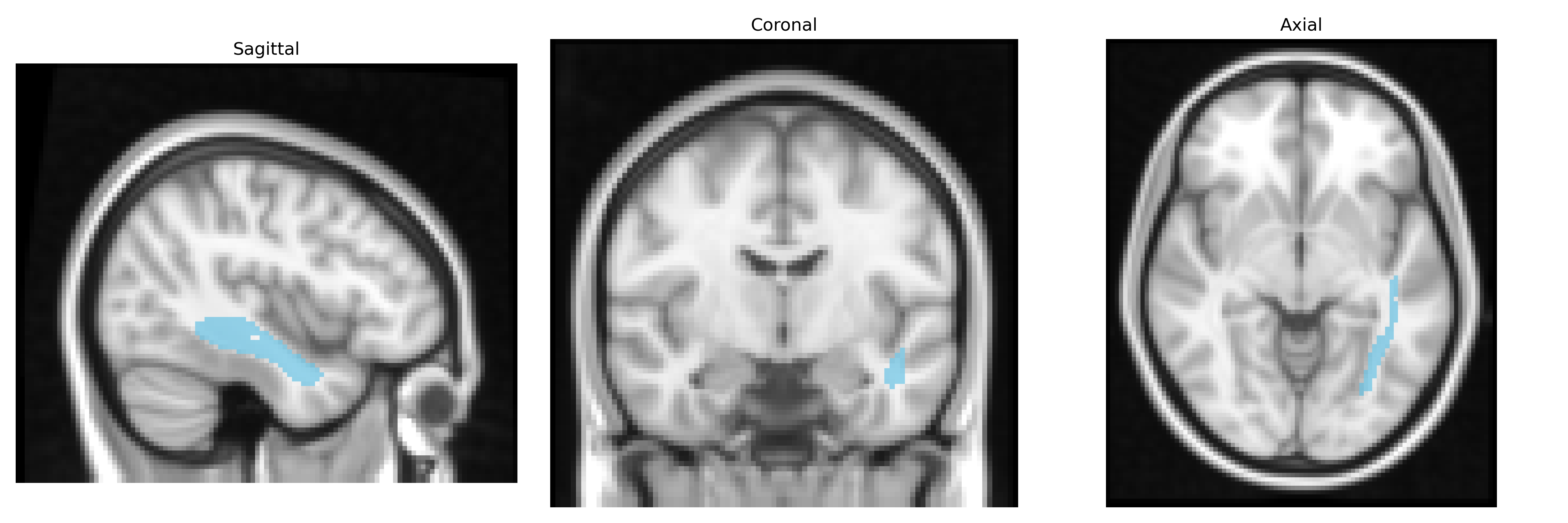
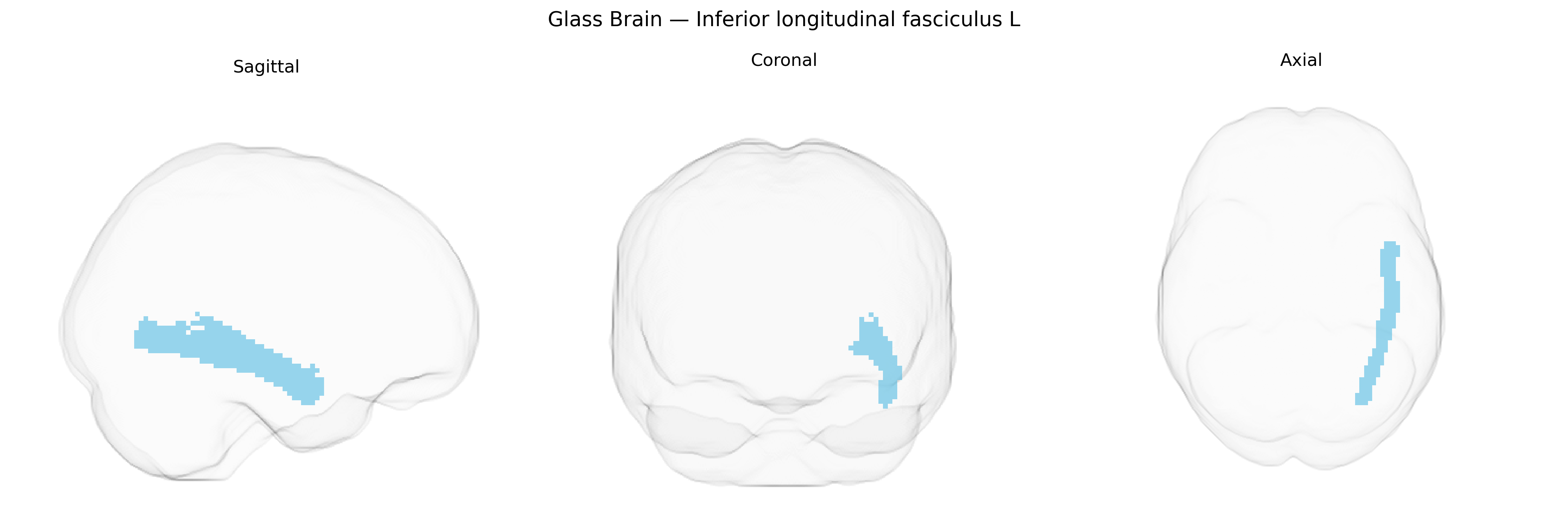
The bilateral Inferior Longitudinal Fasciculus (ILF) in the left hemisphere, as defined in the JHU ICBM DTI-81 white-matter tractography atlas (maxprob thr25, 2 mm), is a major associative fiber bundle that runs longitudinally within the ventral temporal lobe, connecting occipital regions (including visual association cortex) with anterior and medial temporal structures such as the fusiform gyrus, parahippocampal gyrus, and temporal pole. The ILF courses parallel to the ventricular system and below the optic radiations, supporting the integration of high-level visual information with memory, semantic processing, and aspects of emotion and face-object recognition. Functionally, it is implicated in ventral visual stream processing, including object and word recognition, and its disruption has been associated with deficits in visual perception, reading, and certain neuropsychiatric and neurodegenerative conditions. There is no direct Wikipedia article for this specific JHU atlas label; see the related structure Inferior longitudinal fasciculus.
The bilateral inferior longitudinal fasciculus (ILF), as defined in the JHU ICBM tracts maxprob thr25 2mm atlas, has been implicated in several genetic and imaging-genetics studies, although specific locus-level associations are often reported for broader white matter microstructure measures rather than the ILF alone. Twin and family studies indicate substantial heritability of ILF fractional anisotropy (FA) and mean diffusivity (MD), with SNP-heritability analyses from large DTI-GWAS consortia (e.g., ENIGMA, UK Biobank) implicating polygenic influences from genes involved in axonal growth, myelination, and synaptic function, including pathways related to neurodevelopment and cell adhesion (such as those involving NRG1/ErbB signaling, cell-adhesion molecules, and oligodendrocyte-related genes), though specific variants and genes differ across studies and rarely reach genome-wide significance for ILF alone. ILF alterations have been reported in schizophrenia, bipolar disorder, major depression, autism spectrum disorder, dyslexia, and neurodegenerative diseases, and imaging-genetics work has linked common risk variants for these conditions (for example in genes such as ZNF804A, DISC1, and CACNA1C in some cohorts) to white matter integrity changes that include, but are not limited to, the ILF; however, these effects are modest and regionally nonspecific. GWAS of white matter tracts in population cohorts (notably UK Biobank) show that ILF microstructure shares polygenic architecture with cognitive performance, educational attainment, and psychiatric risk, suggesting that genetic factors influencing ILF integrity contribute to individual differences in higher-order visual processing, language-related networks, and vulnerability to psychopathology, but current evidence does not support a concise list of robust, ILF-specific genetic loci and instead points to a distributed, highly polygenic architecture shared across major association and projection tracts.
Overview generated by GPT-4o (2026).
Region ID: 13
Hemisphere: bilateral
Atlas: JHU ICBM tracts maxprob thr25 2mm
Full Quality Version: Download MP4
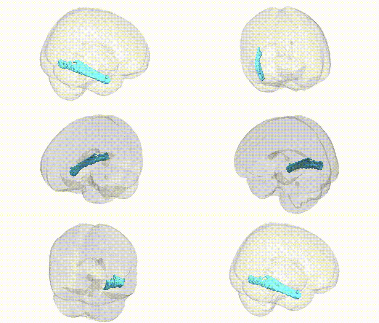
Full Quality Version: Download MP4
Wali Sidiqyar*, Gaurav Rudravaram*, Elyssa M. McMaster, Trent M. Schwartz, Adam M. Saunders, Kurt G. Schilling, Bennett A. Landman "Introducing SPINS: A Shared Public Visualization Library of Neuroanatomical Structures." Medical Imaging with Deep Learning- short paper
This resource is licensed under CC0 1.0 Universal (Public Domain).