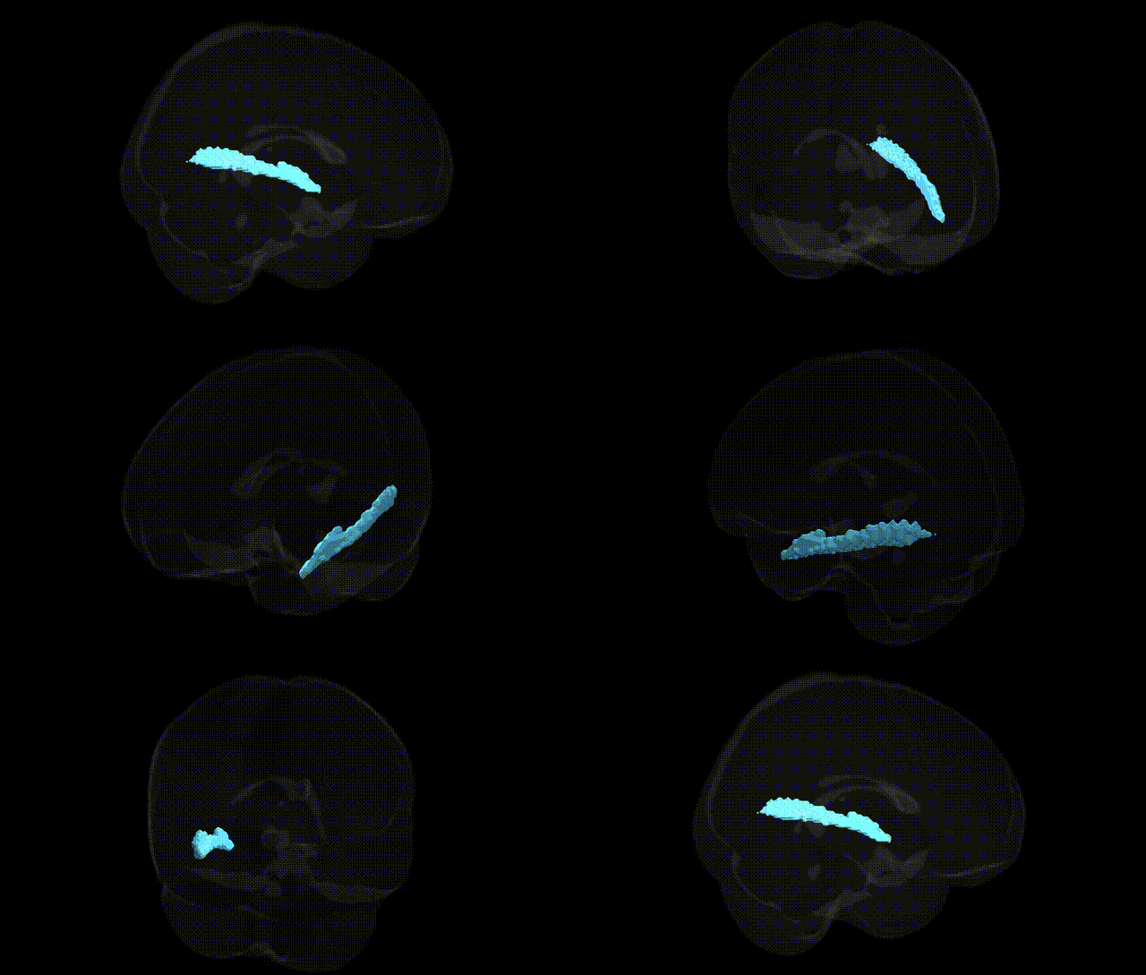
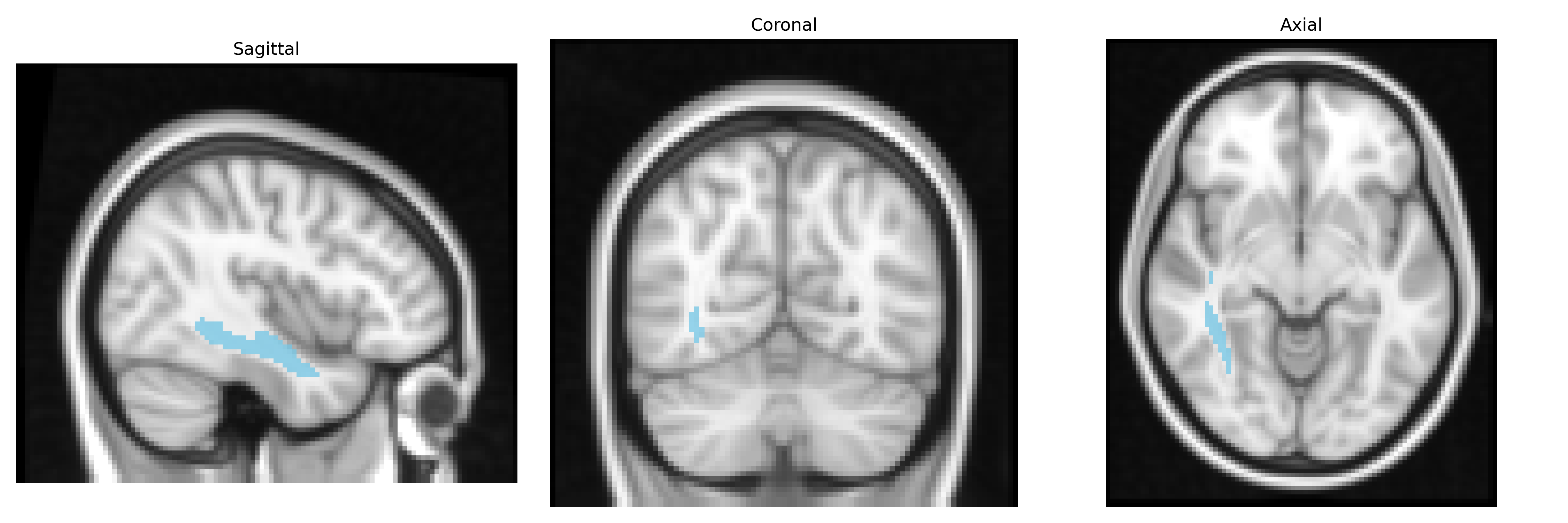
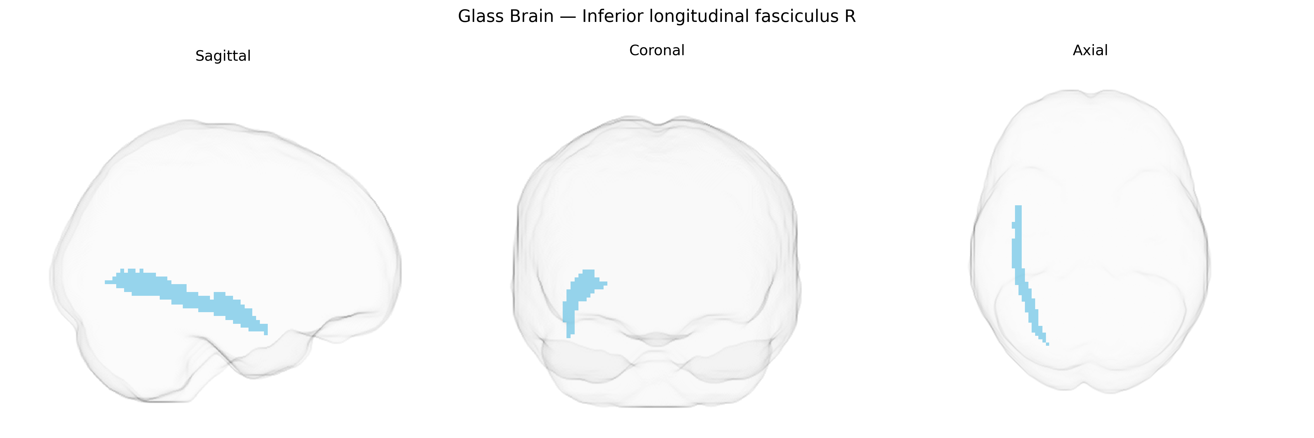
The bilateral Inferior Longitudinal Fasciculus (ILF) is a major associative white matter tract that extends along the ventral temporal and occipital lobes, connecting posterior visual regions in the occipital cortex with anterior temporal areas involved in object recognition, semantic processing, and aspects of emotional perception. In the JHU ICBM tracts maxprob thr25 2mm Atlas, this tract is delineated bilaterally as a long, curved bundle running inferior to the lateral ventricle and temporal horn, integrating visual input with higher-order temporal lobe functions and contributing to processes such as visual object and face recognition, reading, and the integration of visual information with memory and language-related networks. There is no direct link for the Inferior Longitudinal Fasciculus, but a closely related structure is the Temporal lobe.
Genetic associations specifically targeting the bilateral inferior longitudinal fasciculus (ILF), including the right ILF as defined in the JHU ICBM tracts maxprob thr25 2 mm atlas, are relatively sparse, but several imaging–genetics and GWAS studies of white matter microstructure and tract-based features provide indirect evidence. Polygenic influences related to neurodevelopmental and synaptic genes (for example, variants in genes involved in axon guidance, myelination, and neuronal adhesion such as NTRK1/2, CNTN4, and MAG) have been associated with fractional anisotropy and other diffusion MRI metrics in ILF segments, reflecting heritable variation in tract integrity. Large-scale GWAS of white matter tracts (e.g., ENIGMA-DTI and UK Biobank analyses) indicate moderate to high heritability for ILF diffusion measures and implicate loci overlapping pathways for oligodendrocyte function, axonal growth, and neuronal migration, though few loci are uniquely specific to the ILF compared with other association fibers. Genetically influenced alterations of the ILF have been reported in neuropsychiatric and neurodevelopmental conditions such as schizophrenia, autism spectrum disorder, and major depressive disorder, often as part of broader patterns of association fiber disruption; in some cases, disorder-associated risk variants (for example, in genes affecting glutamatergic signaling or neurodevelopment like ZNF804A or DISC1) show imaging correlates that include reduced FA or altered volume in the ILF. Additionally, ILF-related diffusion metrics show polygenic correlations with cognitive traits, including general intelligence and reading ability, and with neuroticism and other personality dimensions, suggesting that shared genetic factors contribute both to ILF microstructure and to behavioral or clinical phenotypes, although no single gene–ILF–trait pathway has yet emerged as definitive.
Overview generated by GPT-4o (2026).
Region ID: 14
Hemisphere: bilateral
Atlas: JHU ICBM tracts maxprob thr25 2mm
Full Quality Version: Download MP4
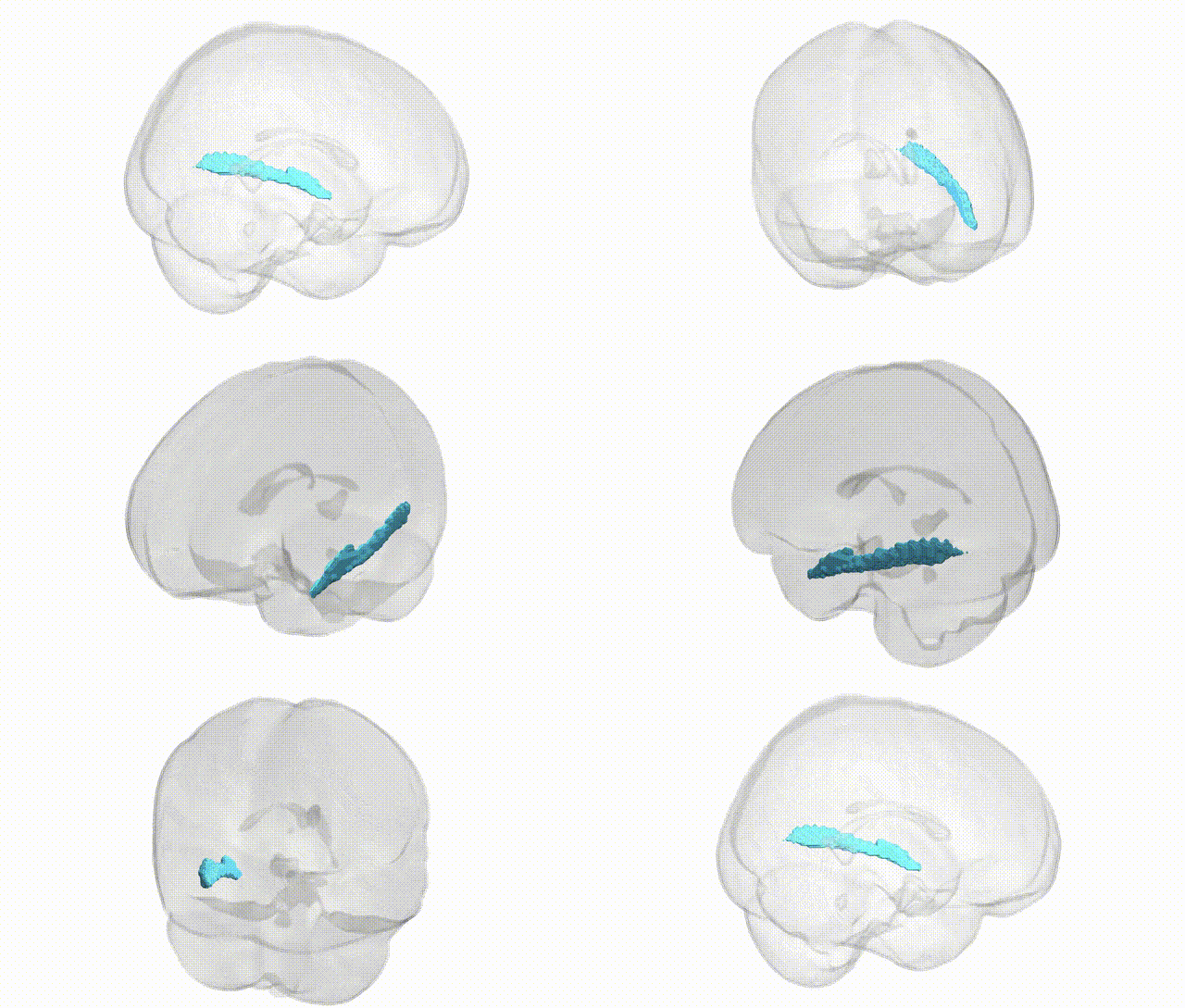
Full Quality Version: Download MP4
Wali Sidiqyar*, Gaurav Rudravaram*, Elyssa M. McMaster, Trent M. Schwartz, Adam M. Saunders, Kurt G. Schilling, Bennett A. Landman "Introducing SPINS: A Shared Public Visualization Library of Neuroanatomical Structures." Medical Imaging with Deep Learning- short paper
This resource is licensed under CC0 1.0 Universal (Public Domain).