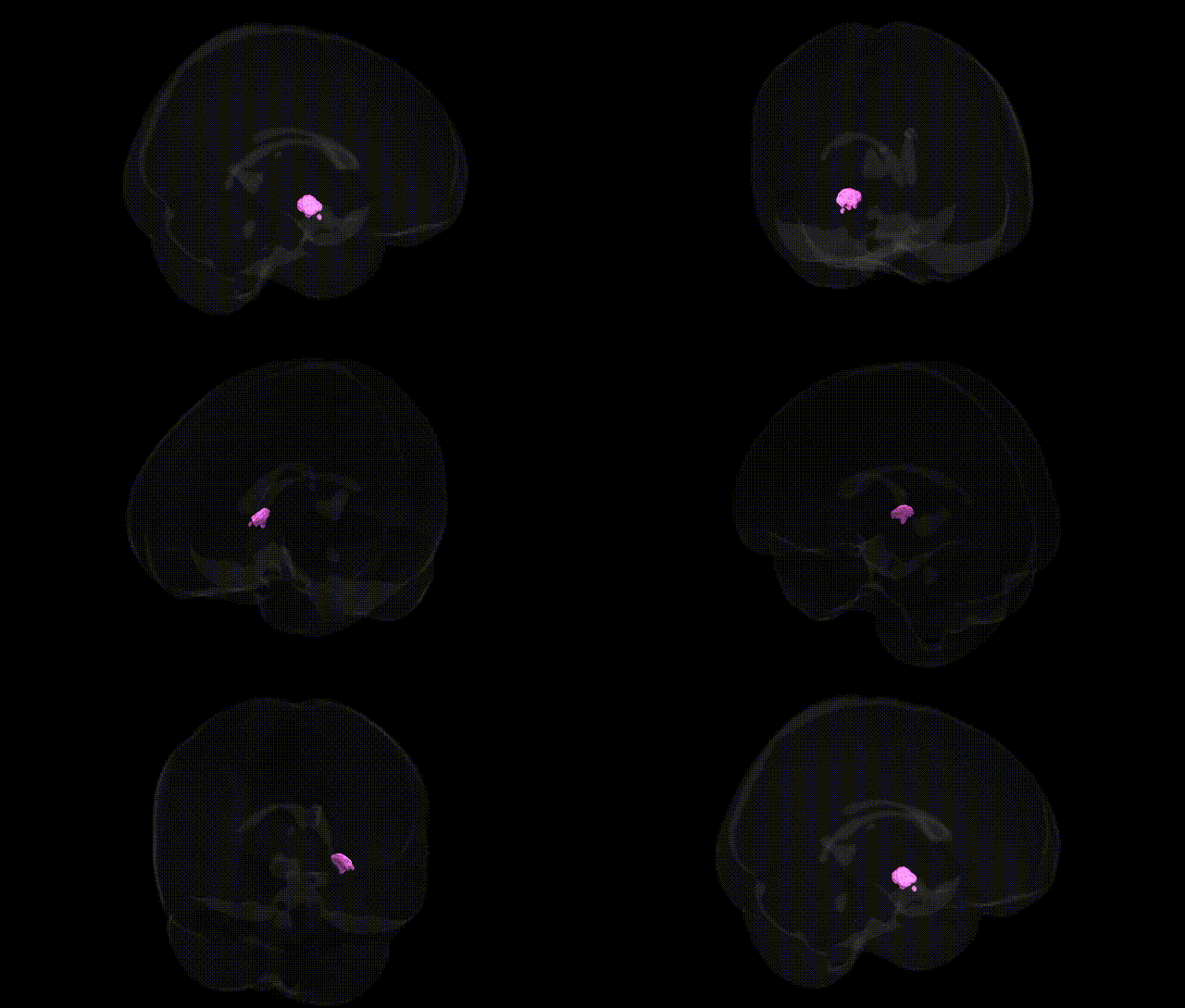
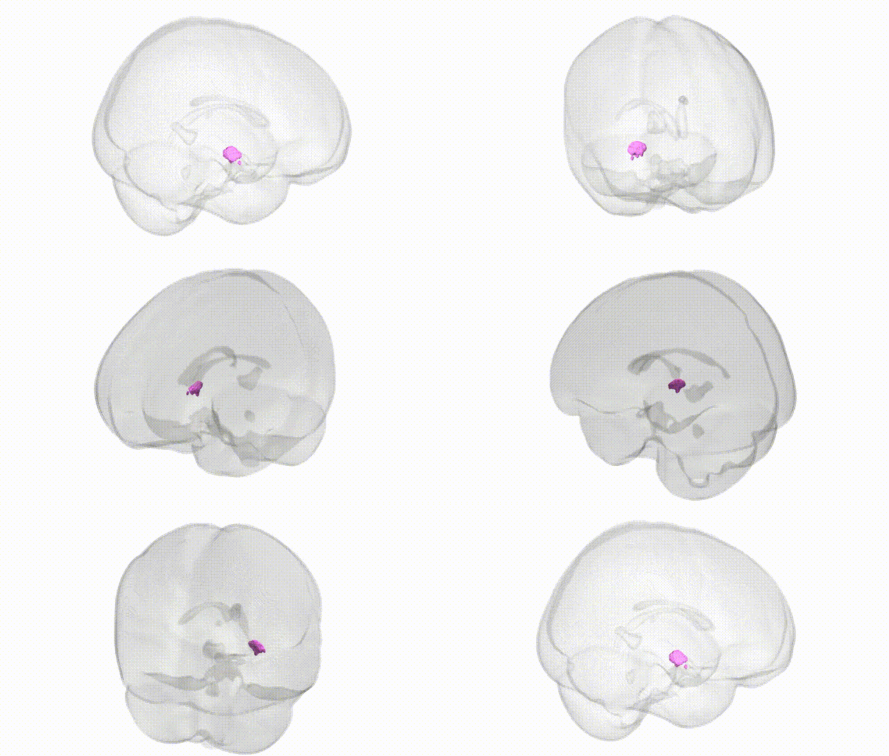
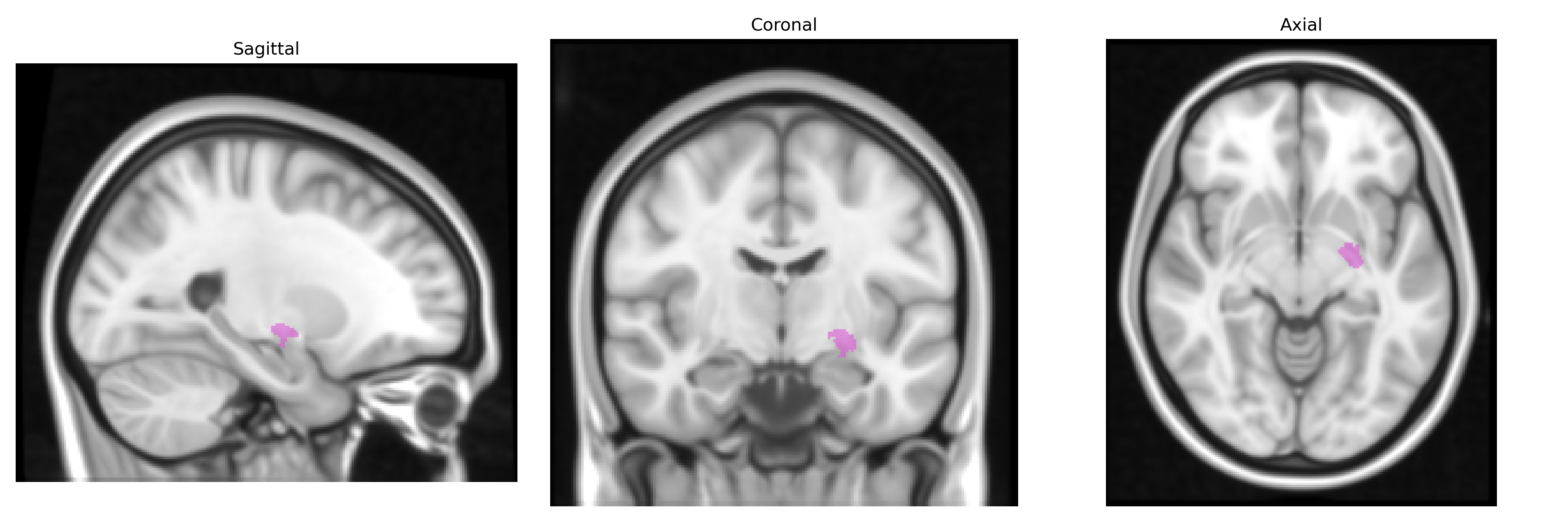
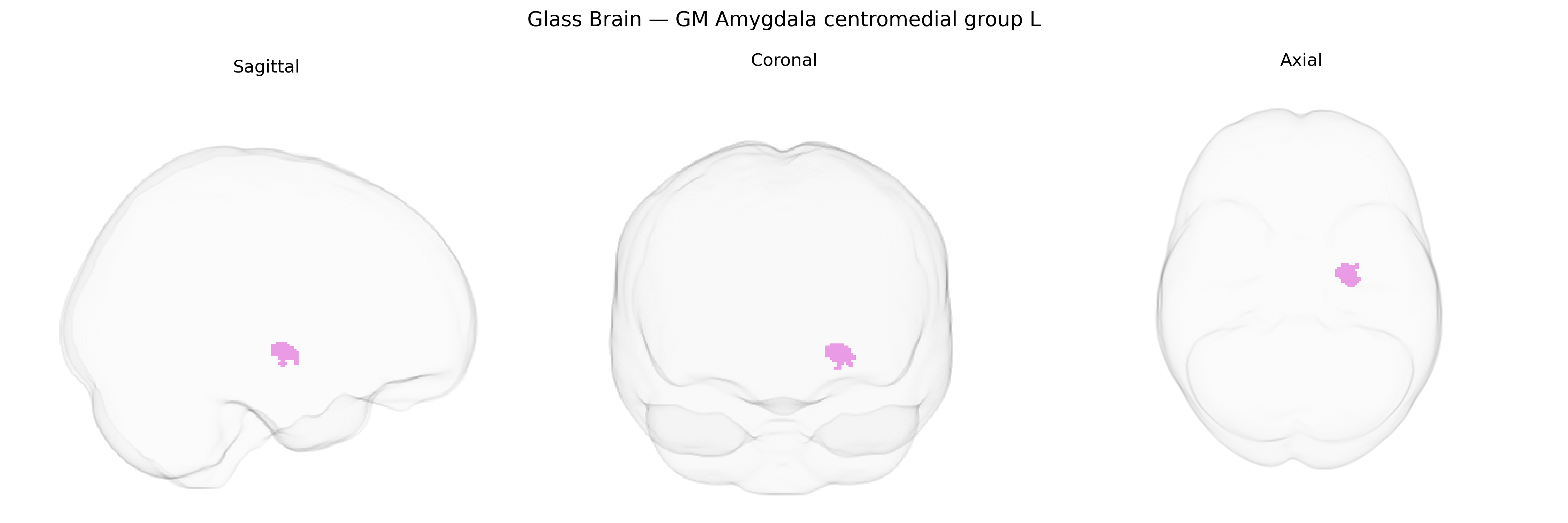
The bilateral GM Amygdala centromedial group L, as defined in the Juelich maxprob thr25 1mm atlas, corresponds to the left centromedial nuclear complex of the amygdala, a set of nuclei located in the medial portion of the temporal lobe’s anterior region. This group is primarily associated with processing emotionally salient stimuli, particularly those related to fear, threat, and social signals, and plays a key role in orchestrating autonomic and behavioral responses via its dense connections to hypothalamic and brainstem centers. Functionally, the centromedial amygdala integrates sensory and limbic inputs and channels them into output patterns that modulate endocrine, cardiovascular, and motor systems, thereby influencing defensive behaviors, stress responses, and aspects of social interaction. Cytoarchitectonically, it is characterized by relatively dense, small neurons and forms part of the broader amygdaloid complex involved in emotional learning and conditioning. There is no direct link for the centromedial group as a distinct region; a related and encompassing structure is the Amygdala.
Genetic associations with the bilateral GM Amygdala centromedial group L (from the Juelich maxprob thr25 1mm atlas) are largely inferred from studies of amygdala volume, functional reactivity, and centromedial subregional mapping rather than this specific parcellation alone, but convergent evidence implicates several gene pathways and traits: common variants in serotonin-related genes (e.g., SLC6A4/5-HTTLPR, HTR1A), glutamatergic and GABAergic signaling genes, and BDNF (notably Val66Met) have been repeatedly associated with altered amygdala structure or hyperreactivity in centromedial regions, particularly in threat-processing and fear-conditioning paradigms; large-scale GWAS of subcortical volumes (e.g., ENIGMA and UK Biobank) have identified multiple loci influencing amygdala volume (including variants near MAD1L1, DRD2, and genes involved in neurodevelopment and synaptic plasticity), though these are typically reported for whole amygdala rather than specific centromedial compartments. Genetically, centromedial amygdala alterations are implicated in anxiety disorders, major depressive disorder, post-traumatic stress disorder, and schizophrenia, with polygenic risk scores for these conditions correlating with amygdala morphology or reactivity; additional associations exist with neuroticism, extraversion, and other personality traits in GWAS linking trait scores to amygdala structure and function. Emerging imaging–genetics work further connects risk loci for autism spectrum disorder and aggression-related traits to centromedial amygdala connectivity, consistent with this region’s role in threat evaluation, emotional salience, and autonomic/endocrine responses.
Overview generated by GPT-4o (2026).
Region ID: 7
Hemisphere: bilateral
Atlas: Juelich maxprob thr25 1mm
Full Quality Version: Download MP4
Full Quality Version: Download MP4
Wali Sidiqyar*, Gaurav Rudravaram*, Elyssa M. McMaster, Trent M. Schwartz, Adam M. Saunders, Kurt G. Schilling, Bennett A. Landman "Introducing SPINS: A Shared Public Visualization Library of Neuroanatomical Structures." Medical Imaging with Deep Learning- short paper
This resource is licensed under CC0 1.0 Universal (Public Domain).