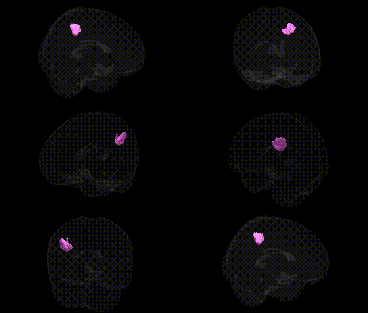
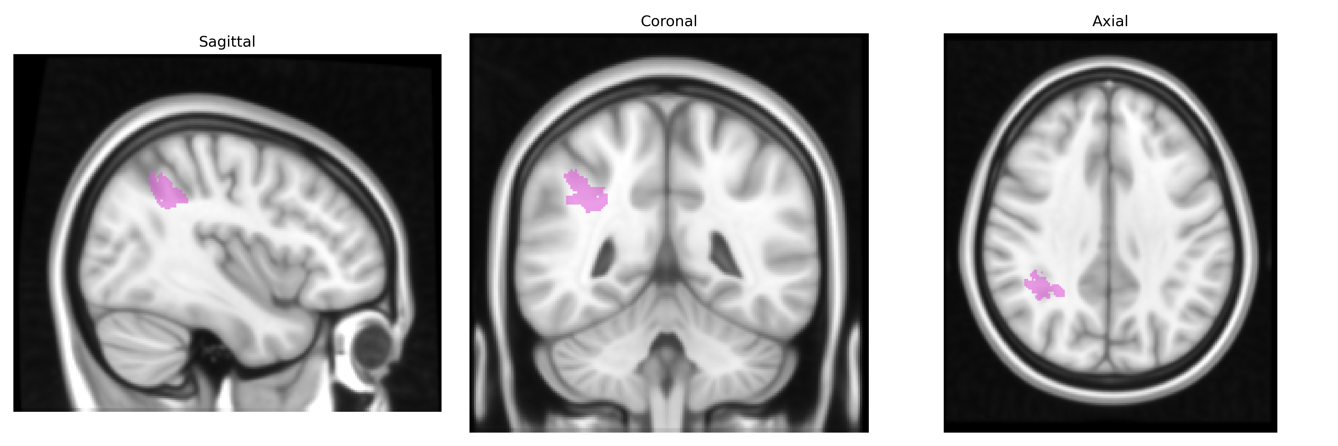
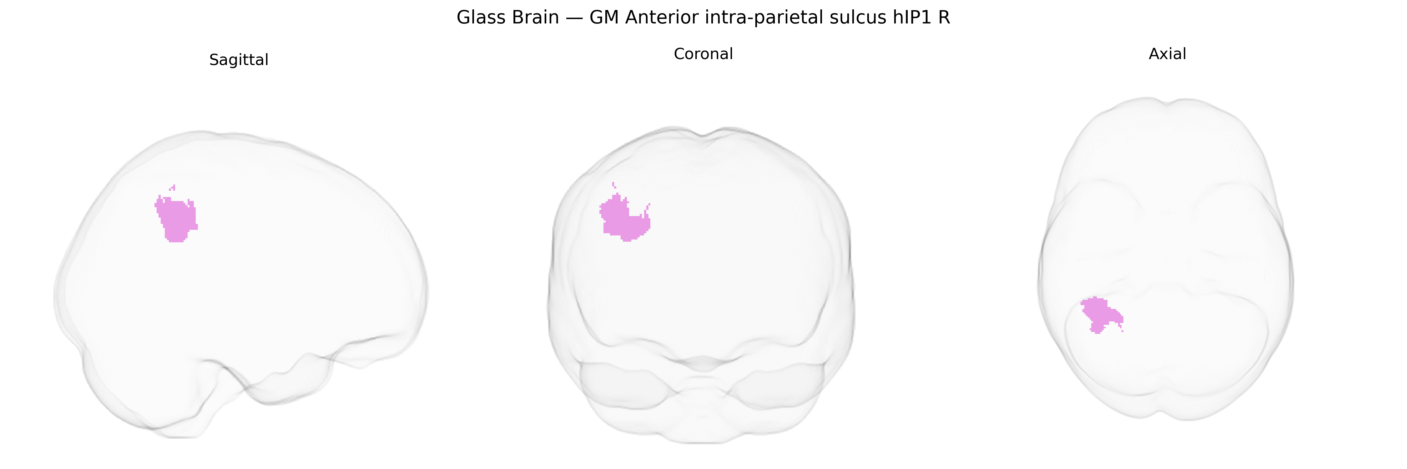
The bilateral GM Anterior intra-parietal sulcus hIP1 R (from the Juelich maxprob thr25 1mm Atlas) refers to a cytoarchitectonically defined right-hemisphere subregion of the anterior intraparietal sulcus within the inferior parietal cortex, implicated in visuomotor integration and object-directed actions such as grasping and hand shaping. This area is thought to participate in transforming visual information about object size, orientation, and spatial position into motor commands, and is closely linked to networks supporting eye–hand coordination, attention to salient visual features, and aspects of sensorimotor decision-making. hIP1 lies within the broader intraparietal sulcus complex, which has multiple functional subdivisions involved in number processing, visuospatial working memory, and spatial attention. There is no direct Wikipedia article for hIP1; a related and encompassing structure is the Intraparietal sulcus.
The bilateral GM anterior intra-parietal sulcus hIP1 (R) region, as defined in the Juelich maxprob atlas, sits within the broader intraparietal sulcus (IPS) circuitry, for which genetic associations have been inferred largely through imaging genetics and GWAS of cortical morphology, cognition, and neuropsychiatric phenotypes rather than region-specific hIP1 data. Large-scale GWAS of cortical thickness and surface area (e.g., ENIGMA, UK Biobank) have implicated common variants near genes involved in neurodevelopment and synaptic function—such as CNP, KIAA0586, genes in Wnt and Notch signaling, and several chromatin regulators—that show associations with structural variation in parietal cortex, including the IPS. Functionally, IPS subregions including hIP1 are central to visuospatial attention, number processing, and sensorimotor integration, and GWAS of mathematical ability, general cognitive performance, and educational attainment have identified polygenic influences (e.g., variants near FOXP2, DCDC2, and other neurodevelopmental genes) whose intermediate phenotypes include IPS activation and structure, though not always specifically hIP1. Neuropsychiatric GWAS in disorders such as ADHD, schizophrenia, and autism have linked risk variants in synaptic and neurodevelopmental genes (e.g., CACNA1C, GRIN2A, CNTNAP2) to altered parietal activation or morphology, with IPS involvement repeatedly observed in case–control imaging studies; however, these links remain indirect and regionally coarse rather than establishing unique genetic specificity for hIP1. Overall, current evidence suggests that genetic influences on hIP1 mirror those on the larger IPS–parietal network, with polygenic contributions to cortical development, cognitive abilities, and psychiatric risk shaping structure and function, but no robust GWAS yet isolates hIP1 as a distinct target with specific variants uniquely associated to this atlas-defined area.
Overview generated by GPT-4o (2026).
Region ID: 2
Hemisphere: bilateral
Atlas: Juelich maxprob thr25 1mm
Full Quality Version: Download MP4
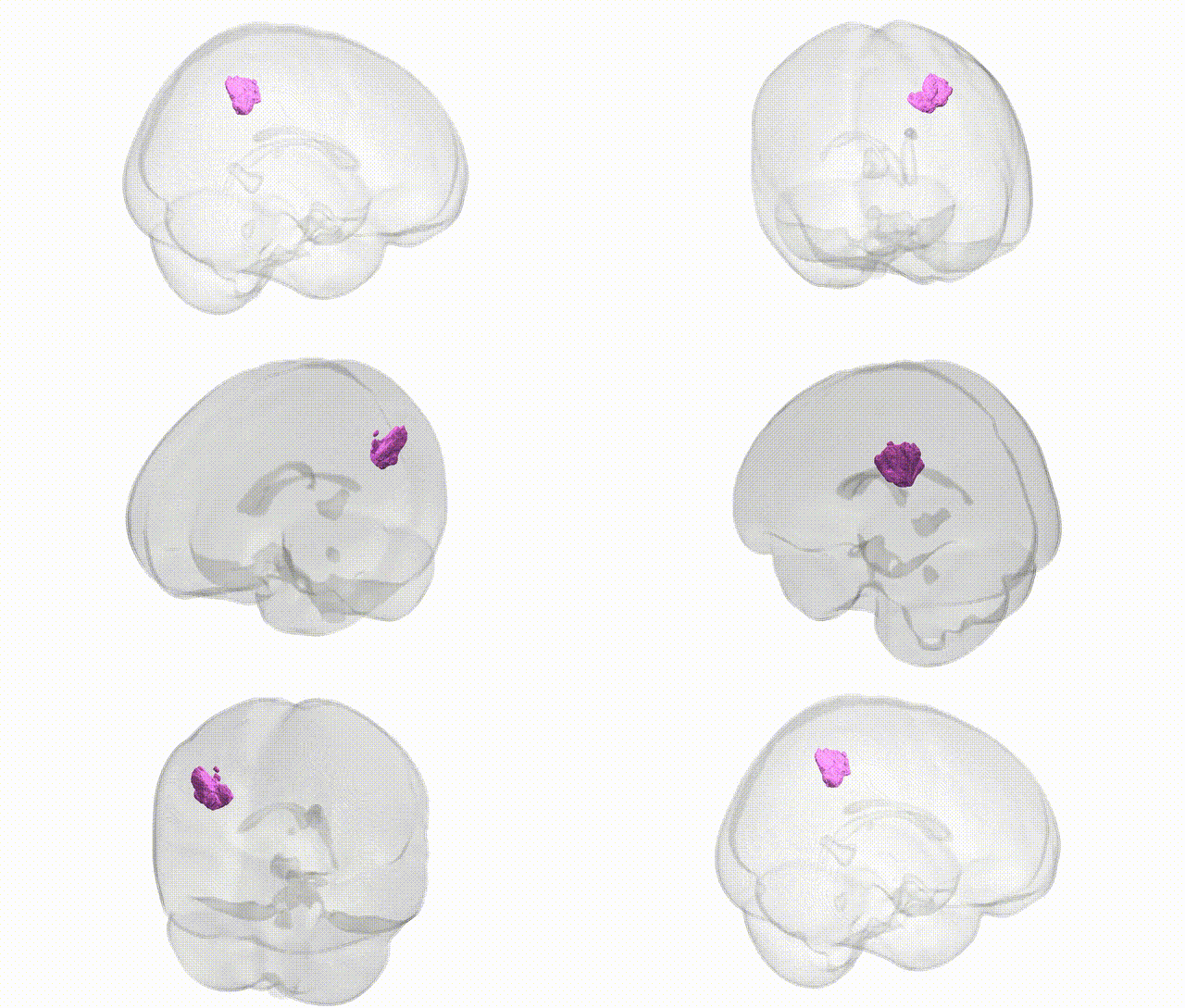
Full Quality Version: Download MP4
Wali Sidiqyar*, Gaurav Rudravaram*, Elyssa M. McMaster, Trent M. Schwartz, Adam M. Saunders, Kurt G. Schilling, Bennett A. Landman "Introducing SPINS: A Shared Public Visualization Library of Neuroanatomical Structures." Medical Imaging with Deep Learning- short paper
This resource is licensed under CC0 1.0 Universal (Public Domain).