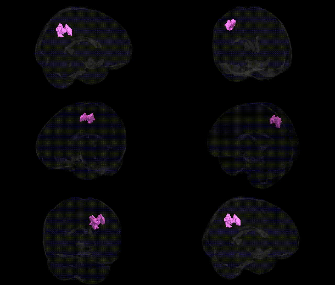
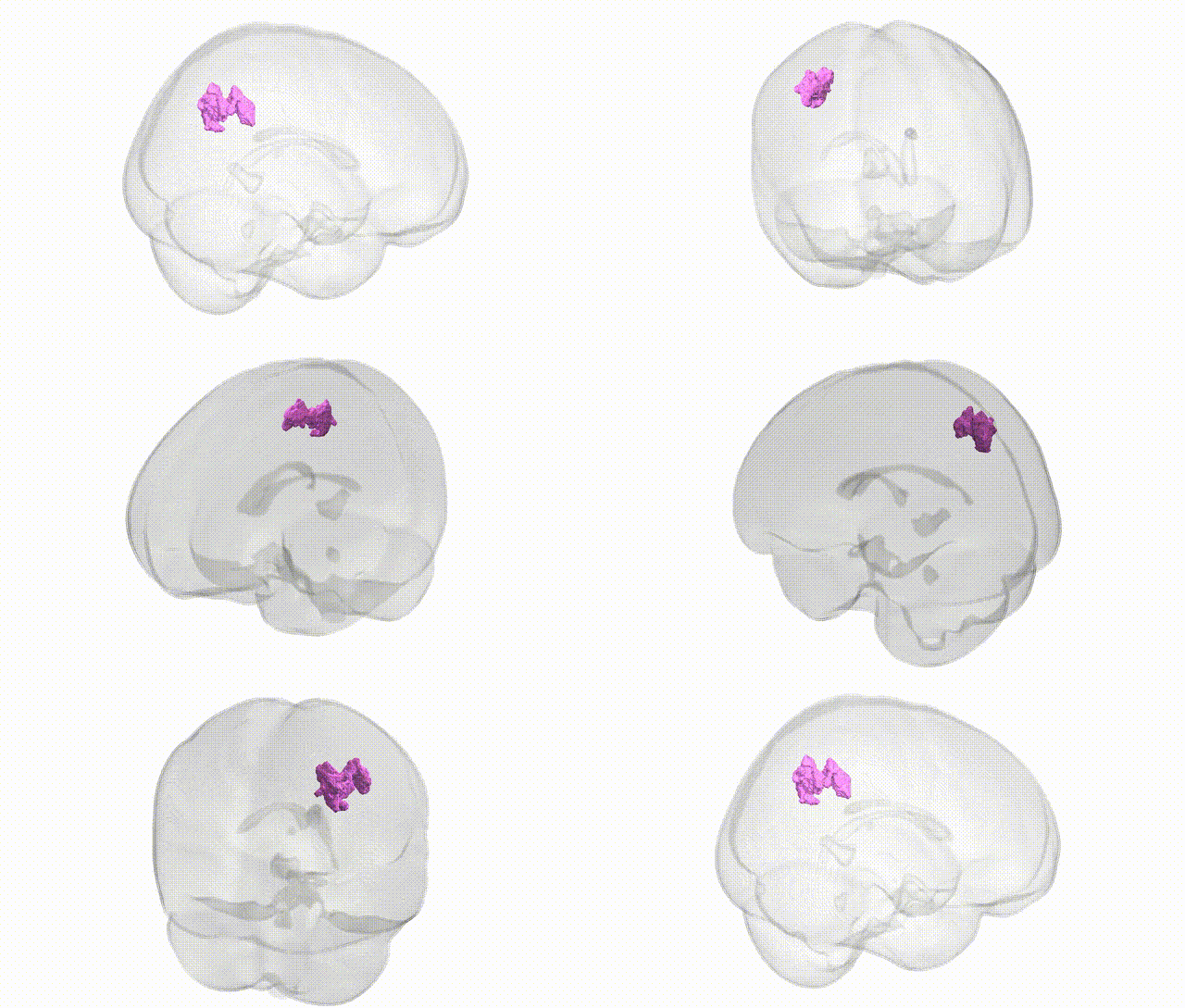
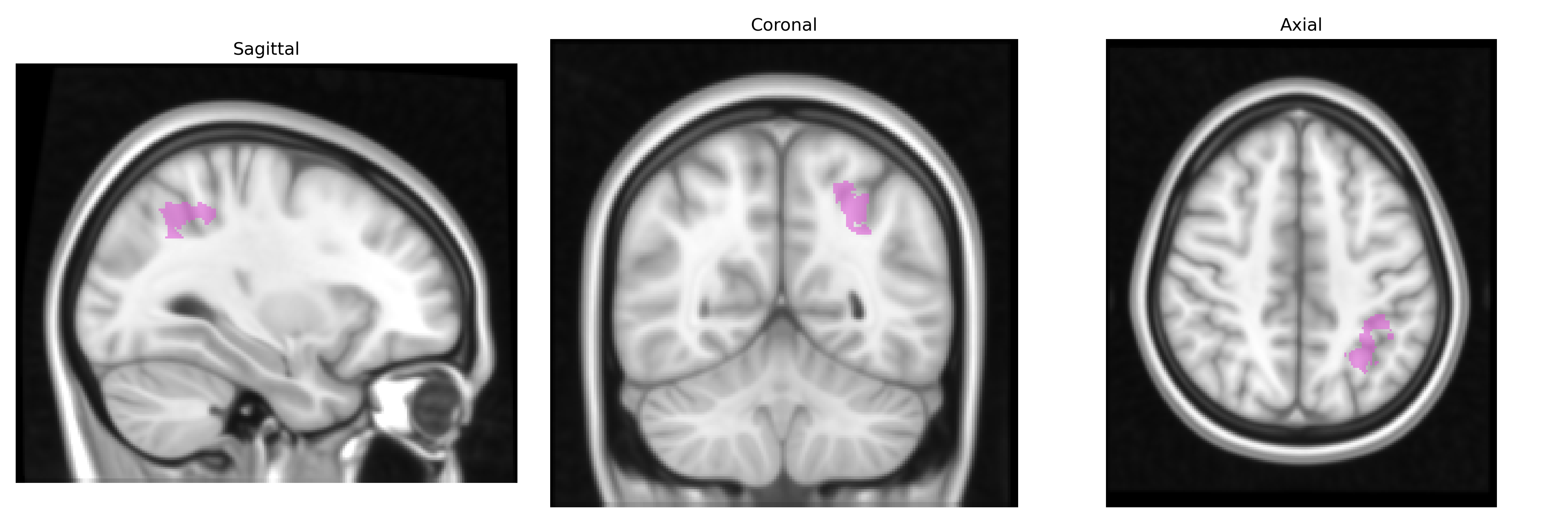
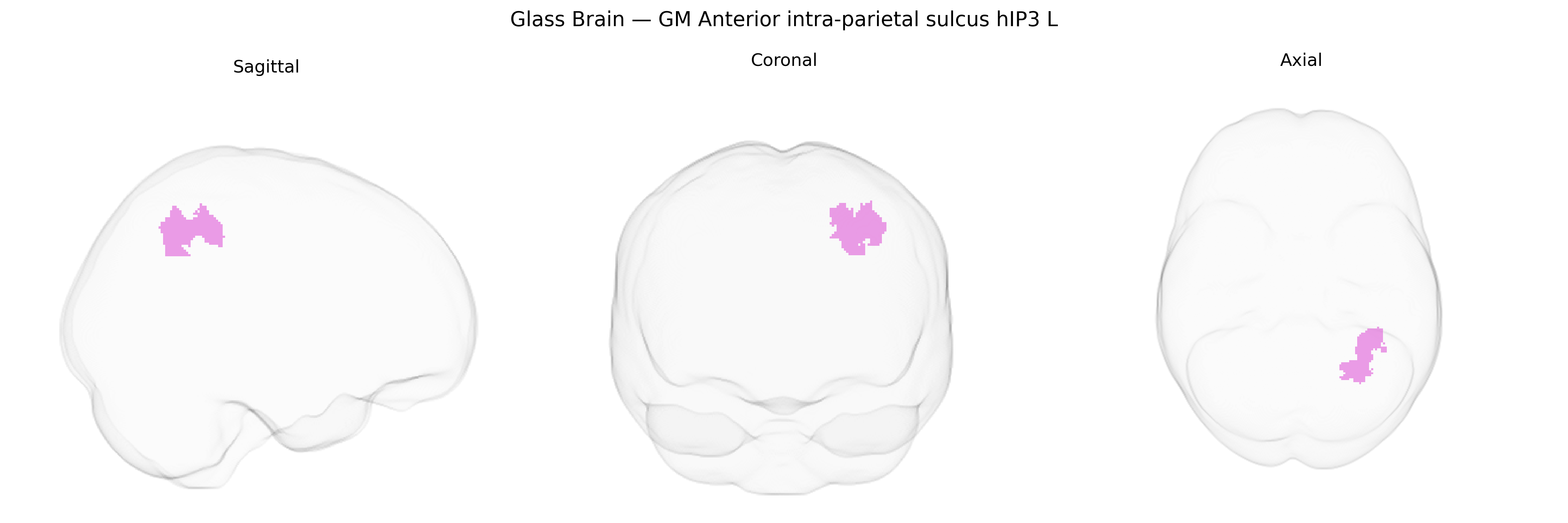
The bilateral GM Anterior intra-parietal sulcus hIP3 L region, defined in the Juelich maxprob thr25 1 mm atlas, corresponds to a cytoarchitectonically and functionally characterized subregion within the anterior portion of the intraparietal sulcus of the left hemisphere. This area is part of the posterior parietal cortex and is implicated in visuomotor integration, object-directed actions, and higher-order sensorimotor transformations, including the coordination of hand and eye movements for grasping and manipulation. Neuronal populations within hIP3 exhibit sensitivity to spatial properties of visual stimuli and the planning of goal-directed movements, integrating visual, somatosensory, and motor-related signals to support fine motor control and interaction with objects in extrapersonal space. There is no dedicated Wikipedia page for hIP3; a closely related and encompassing structure is the Intraparietal sulcus.
The anterior intraparietal sulcus area hIP3, a higher-order visuomotor and attention-related region in the Juelich atlas, has been implicated indirectly in genetic studies through its role in frontoparietal and dorsal attention networks rather than through region-specific GWAS hits. Large neuroimaging genetics consortia (e.g., ENIGMA, UK Biobank) have identified numerous common variants (including loci near genes such as HMGA2, KIAA0586, and others involved in neurodevelopment and synaptic function) associated with parietal cortical thickness and surface area, which encompass intraparietal sulcus territories like hIP3. GWAS of cognitive performance, working memory, intelligence, and educational attainment—traits that robustly engage the intraparietal sulcus functionally—have highlighted polygenic architectures involving neurodevelopmental and synaptic genes (e.g., variants near MAPT, DCC, and multiple glutamatergic and GABAergic pathway genes), with imaging-genetics work showing that such variants modulate activation and connectivity within frontoparietal networks including AIP/hIP regions. Disorder-focused genetic studies in attention-deficit/hyperactivity disorder, schizophrenia, and autism spectrum disorder frequently implicate genes affecting parietal cortex development and connectivity (such as those in chromatin remodeling, axon guidance, and calcium channel pathways), and case–control imaging-genetics analyses often find altered structure or function in the intraparietal sulcus that covaries with polygenic risk for these conditions. However, no well-established GWAS signals are currently known that specifically and uniquely localize to bilateral GM hIP3 as defined by the Juelich maxprob thr25 1 mm atlas; rather, genetic associations are expressed at the level of distributed parietal and frontoparietal systems that include hIP3 as a key node.
Overview generated by GPT-4o (2026).
Region ID: 5
Hemisphere: bilateral
Atlas: Juelich maxprob thr25 1mm
Full Quality Version: Download MP4
Full Quality Version: Download MP4
Wali Sidiqyar*, Gaurav Rudravaram*, Elyssa M. McMaster, Trent M. Schwartz, Adam M. Saunders, Kurt G. Schilling, Bennett A. Landman "Introducing SPINS: A Shared Public Visualization Library of Neuroanatomical Structures." Medical Imaging with Deep Learning- short paper
This resource is licensed under CC0 1.0 Universal (Public Domain).