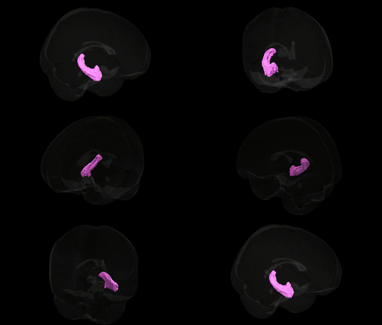
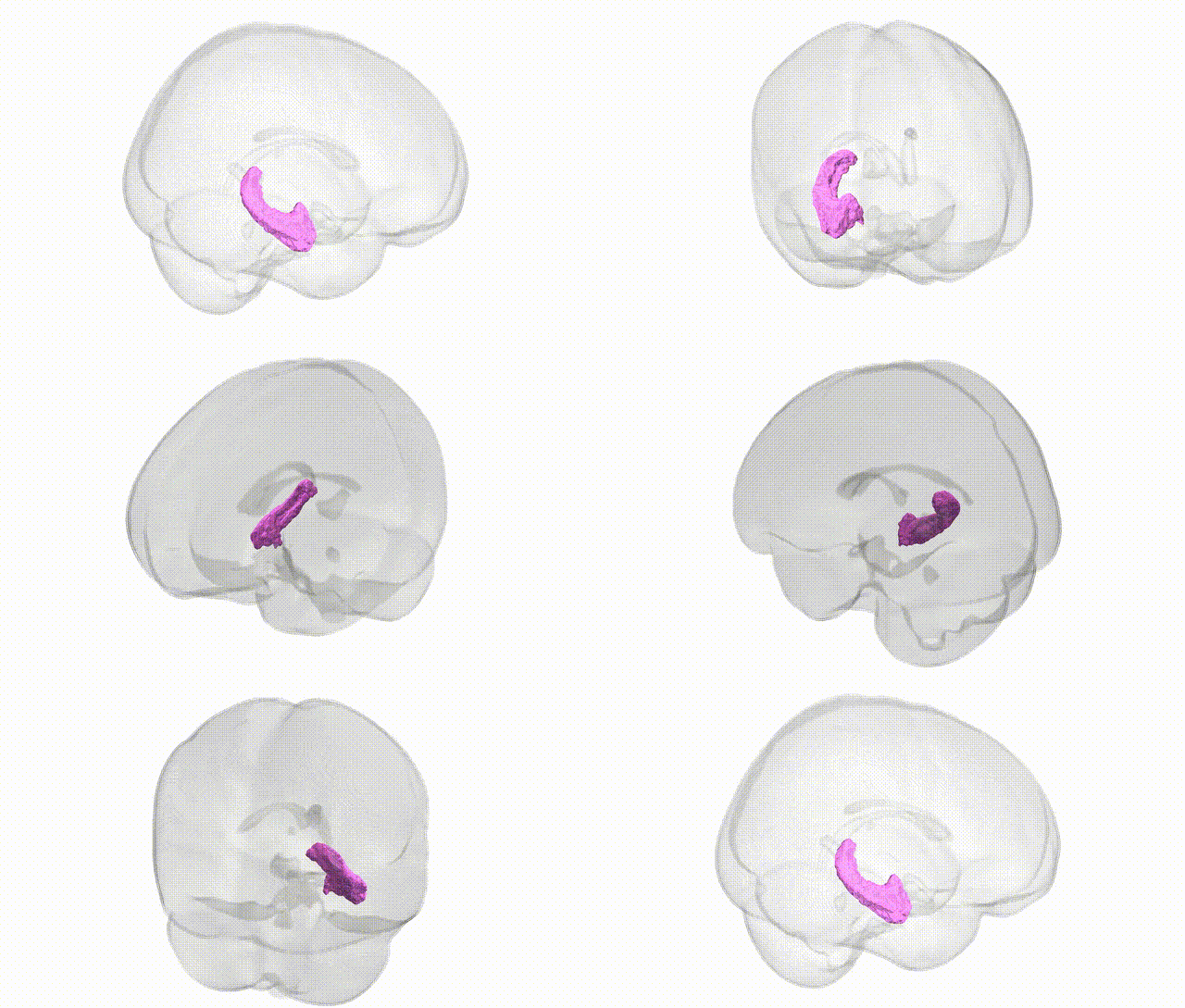
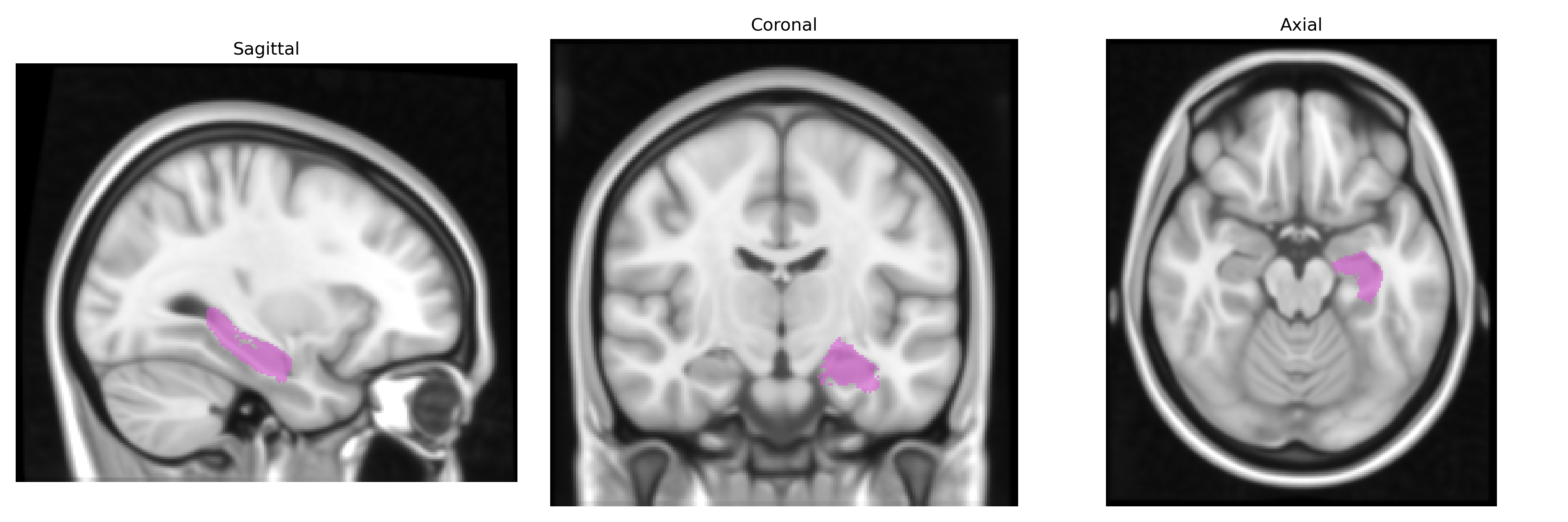
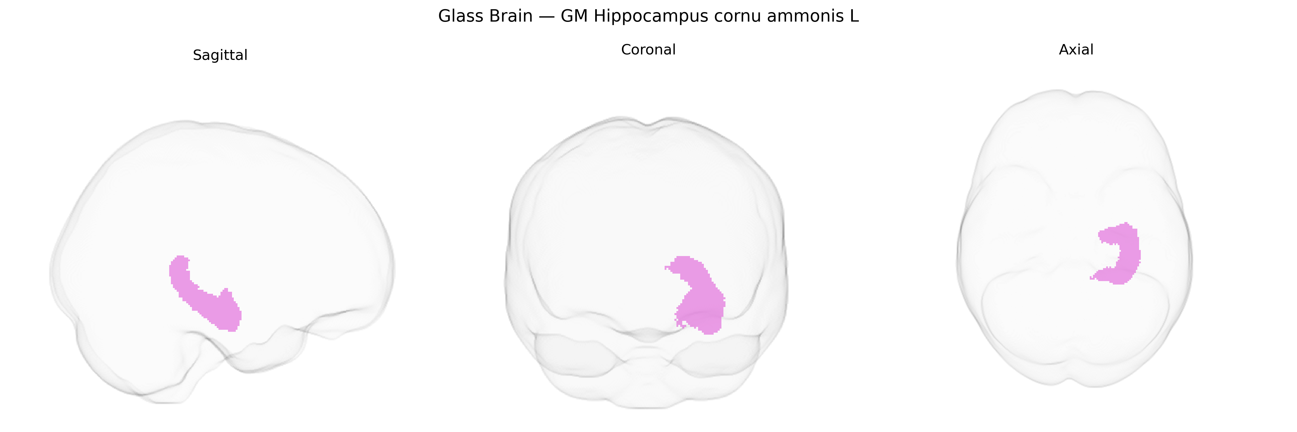
The bilateral GM Hippocampus cornu ammonis L, as defined in the Juelich maxprob thr25 1 mm atlas, refers to the gray matter of the left cornu ammonis (CA) fields of the hippocampus, a key medial temporal lobe structure involved in episodic memory formation, spatial navigation, and contextual learning. Histologically, the cornu ammonis comprises distinct subfields (CA1–CA4) characterized by densely packed pyramidal neurons, each subfield differing in cytoarchitecture, connectivity, and vulnerability to neuropathological processes such as hypoxia and epilepsy. Functionally, the CA regions integrate multimodal cortical input via the entorhinal cortex and contribute to pattern separation and completion through recurrent collateral networks and interactions with the dentate gyrus and subiculum. In humans, this region is crucial for encoding and retrieval of declarative memories and is a primary locus of early degeneration in disorders such as Alzheimer’s disease. There is no direct Wikipedia article for “Hippocampus cornu ammonis”; a closely related and encompassing structure is the hippocampus: Hippocampus.
The bilateral gray matter hippocampal cornu ammonis (CA) subfields in the Juelich maxprob thr25 1mm atlas are key components of hippocampal circuitry, and GWAS of hippocampal volume and subfield morphology have identified multiple genetic associations, notably in and around genes such as DPP4, ASTN2, MSRB3, HRK, WWOX, and variants near APOE, which influence overall hippocampal integrity and age-related atrophy. Large-scale imaging genetics studies (e.g., ENIGMA) have shown that common variants affecting glutamatergic signaling, neurodevelopment, and synaptic plasticity are associated with CA subfield volumes, supporting a role in memory and learning traits. Polygenic risk for Alzheimer’s disease, particularly driven by APOE and other AD loci (CLU, PICALM, BIN1), has been robustly linked to reduced CA subfield volume and accelerated atrophy, and similar though generally weaker associations have been reported for schizophrenia, major depressive disorder, and bipolar disorder, where risk variants in genes such as CACNA1C, GRM3, and ZNF804A correlate with hippocampal abnormalities. GWAS of cognitive performance, educational attainment, and general intelligence show overlapping genetic architecture with hippocampal CA volume, indicating pleiotropy between genes influencing these traits and those shaping hippocampal microstructure. Although most studies consider total hippocampal or broad subfield volumes rather than the specific Juelich-defined bilateral GM CA L region, convergent evidence implies that the genetic variants modulating hippocampal neurogenesis, synaptic function, and vulnerability to neurodegeneration are also relevant to this CA territory and its contributions to episodic memory, stress regulation, and disease risk.
Overview generated by GPT-4o (2026).
Region ID: 17
Hemisphere: bilateral
Atlas: Juelich maxprob thr25 1mm
Full Quality Version: Download MP4
Full Quality Version: Download MP4
Wali Sidiqyar*, Gaurav Rudravaram*, Elyssa M. McMaster, Trent M. Schwartz, Adam M. Saunders, Kurt G. Schilling, Bennett A. Landman "Introducing SPINS: A Shared Public Visualization Library of Neuroanatomical Structures." Medical Imaging with Deep Learning- short paper
This resource is licensed under CC0 1.0 Universal (Public Domain).