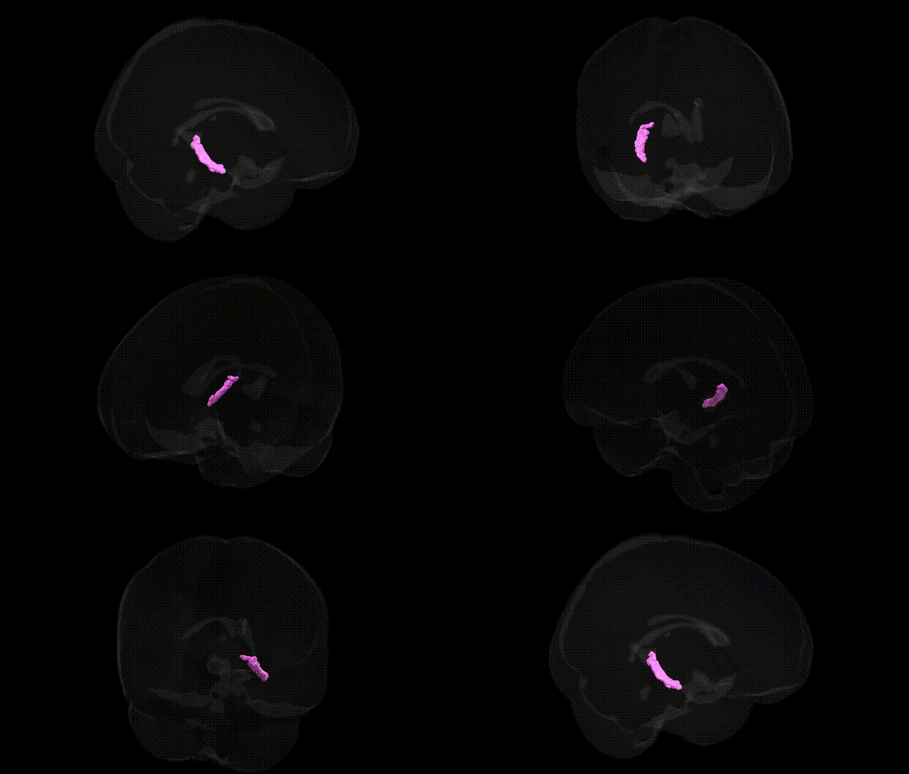
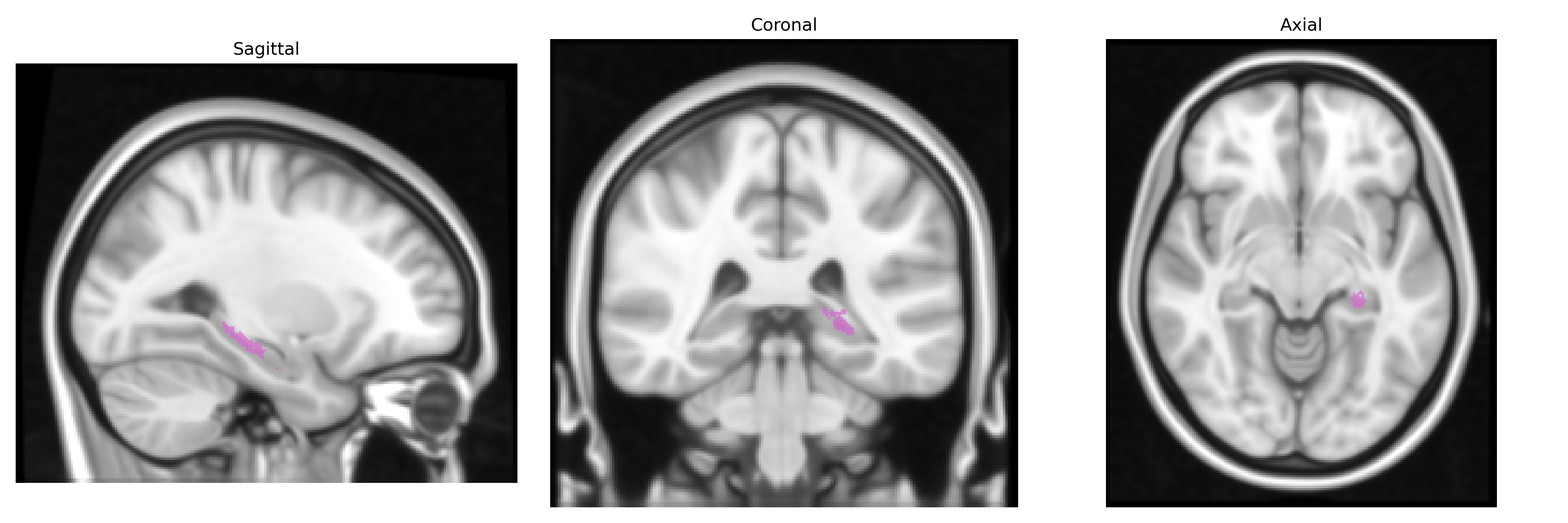
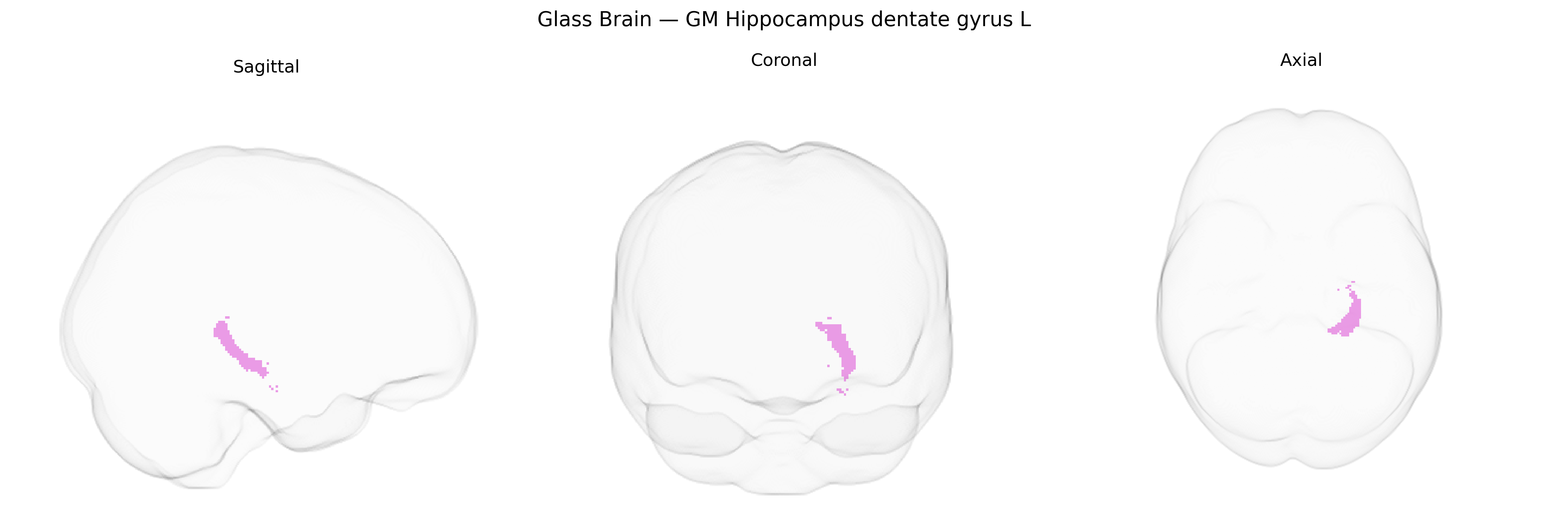
The bilateral GM Hippocampus dentate gyrus L, as defined in the Juelich maxprob thr25 1mm atlas, corresponds to the left gray-matter portion of the dentate gyrus, a key subregion of the hippocampal formation situated in the medial temporal lobe. This structure is characterized by its densely packed granule cells, which receive excitatory input primarily from the entorhinal cortex via the perforant path and project to the CA3 region of the hippocampus, forming the first stage of the classic trisynaptic circuit. Functionally, the dentate gyrus is strongly implicated in pattern separation, episodic memory encoding, and aspects of spatial navigation, and it remains one of the few regions in the adult mammalian brain where adult neurogenesis has been robustly documented. Although no dedicated Wikipedia entry exists for this specific Juelich-defined region, it is encompassed within the broader hippocampal formation described here: Hippocampus.
Genetic associations involving the bilateral hippocampal dentate gyrus, including its left Juelich-defined subfield, converge on pathways for neurodevelopment, synaptic plasticity, and stress response, with multiple GWAS linking common variants to hippocampal and dentate gyrus volume, pattern separation–related cognition, and risk for psychiatric and neurodegenerative disorders. Large-scale imaging-genetics studies (e.g., ENIGMA, UK Biobank) have identified loci near or within genes such as BDNF, MAPT, SLC4A10, WIF1, DPP4, and several chromatin-remodeling and axon-guidance genes that influence hippocampal subfield volumes, including dentate gyrus–enriched regions, and show partial overlap with Alzheimer’s disease, schizophrenia, major depression, and bipolar disorder risk loci. Schizophrenia and major depressive disorder GWAS highlight polygenic overlap with hippocampal structure, implicating genes involved in glutamatergic signaling, GABAergic interneuron function, and neurogenesis, consistent with dentate gyrus vulnerability in these conditions. Alzheimer’s disease risk variants (e.g., in APOE, BIN1, CLU, PICALM) correlate with reduced hippocampal volume and altered subfield integrity, including the dentate gyrus, and are associated with memory decline trajectories. Additional associations link stress-related genes (e.g., FKBP5, NR3C1 pathways) and neurotrophin signaling to dentate gyrus–dependent functions such as adult neurogenesis and pattern separation, while polygenic scores for educational attainment, general cognitive ability, and neuroticism show modest but significant relationships with hippocampal subfield structure, further implicating the left dentate gyrus as a genetically modulated node at the intersection of cognition, mood regulation, and neurodegeneration.
Overview generated by GPT-4o (2026).
Region ID: 21
Hemisphere: bilateral
Atlas: Juelich maxprob thr25 1mm
Full Quality Version: Download MP4
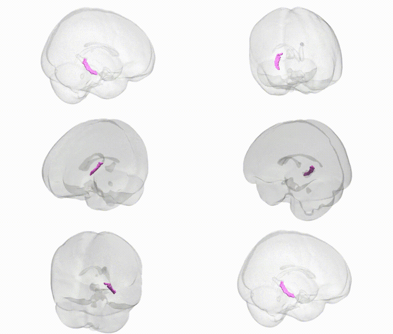
Full Quality Version: Download MP4
Wali Sidiqyar*, Gaurav Rudravaram*, Elyssa M. McMaster, Trent M. Schwartz, Adam M. Saunders, Kurt G. Schilling, Bennett A. Landman "Introducing SPINS: A Shared Public Visualization Library of Neuroanatomical Structures." Medical Imaging with Deep Learning- short paper
This resource is licensed under CC0 1.0 Universal (Public Domain).