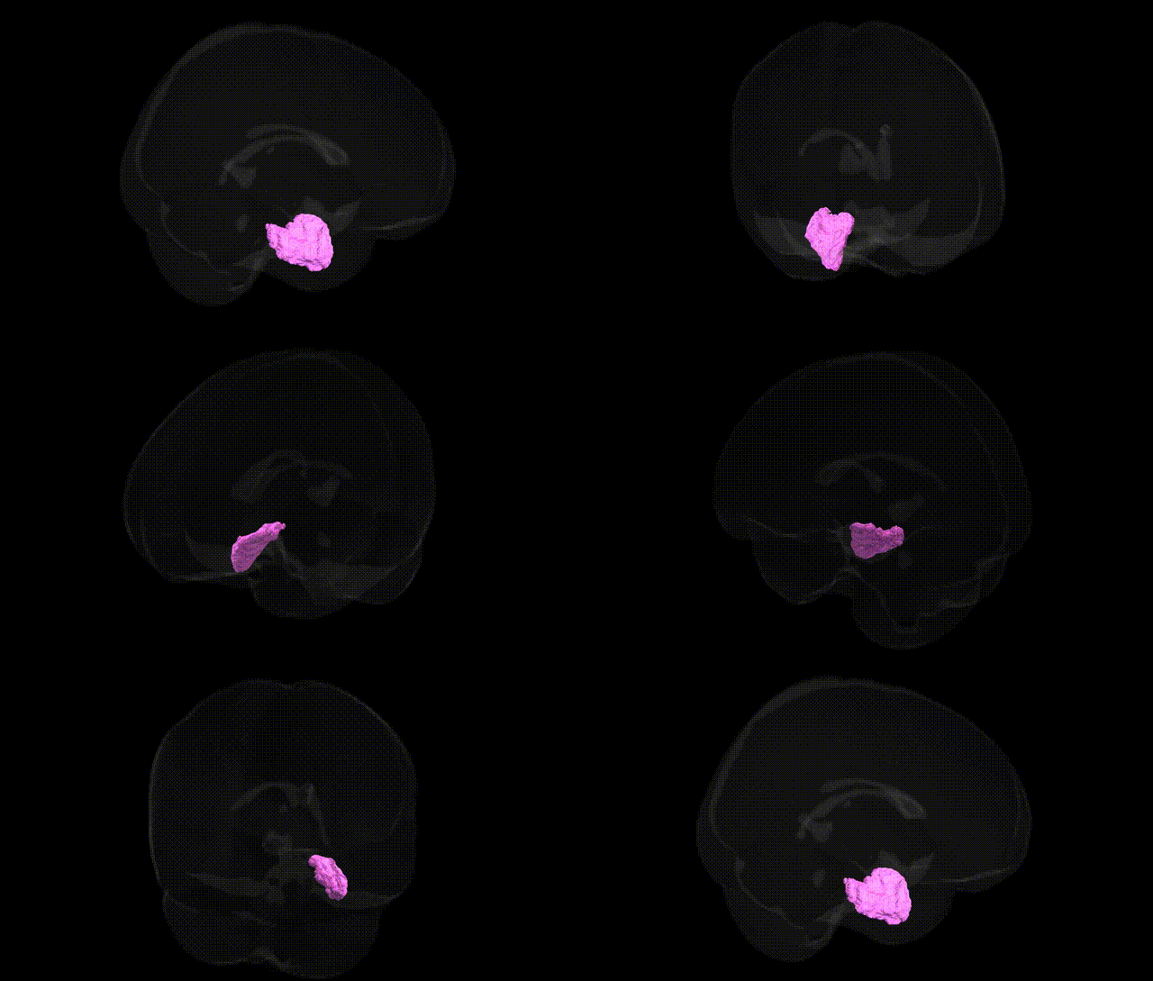
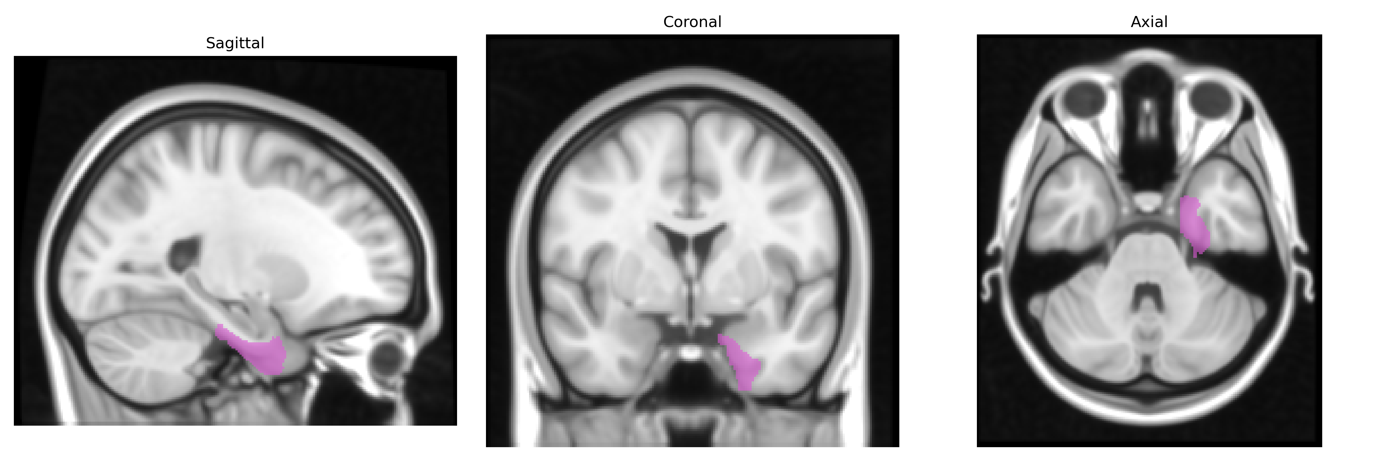
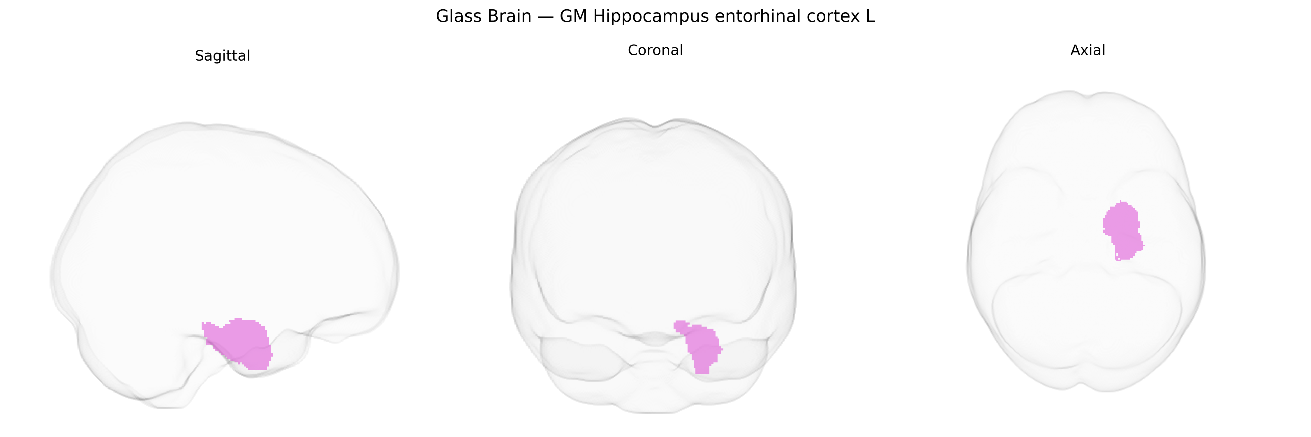
The bilateral GM Hippocampus entorhinal cortex L region in the Juelich maxprob thr25 1mm atlas corresponds primarily to gray matter of the left entorhinal cortex and adjacent hippocampal formation, including portions of the parahippocampal gyrus and hippocampal head and body. This area forms a key interface between neocortex and hippocampus, receiving widespread multimodal cortical input and projecting to hippocampal subfields, particularly the dentate gyrus and CA regions, thereby supporting episodic memory encoding, spatial navigation, and associative learning. Cytoarchitectonically, it is characterized by a laminated cortex with distinct layer II and layer III neuron populations forming the perforant pathway to the hippocampus, and it participates in medial temporal lobe networks critical for declarative memory and early pathological involvement in neurodegenerative diseases such as Alzheimer’s disease. No direct Wikipedia article exists for this exact combined label; a related article is available for the Entorhinal cortex.
The left entorhinal cortex (bilateral GM Hippocampus entorhinal cortex L in the Juelich maxprob thr25 1mm atlas) shows robust genetic associations with Alzheimer’s disease (AD) risk and related endophenotypes, as numerous GWAS and imaging–genetics studies identify variants in and around APOE (especially ε4), BIN1, CLU, ABCA7, SORL1, and PICALM as influencing entorhinal and hippocampal volume, cortical thickness, and neurodegeneration, with APOE-ε4 carriers typically showing early entorhinal atrophy. Polygenic risk scores for AD and general cognitive ability also correlate with entorhinal thickness and hippocampal–entorhinal structural integrity, while large-scale ENIGMA and UK Biobank analyses implicate common variants in genes related to lipid metabolism, synaptic function, and neurodevelopment (e.g., WWOX, KIBRA/WWC1, MAPT region) in interindividual variability of medial temporal lobe morphology. Beyond AD, genetic risk for temporal lobe epilepsy, frontotemporal dementia, and schizophrenia has been associated with alterations in entorhinal and adjacent hippocampal structures, and loci affecting general brain morphometry, such as those near HMGA2 and IGF1, contribute to overall medial temporal volume differences that include the left entorhinal cortex.
Overview generated by GPT-4o (2026).
Region ID: 19
Hemisphere: bilateral
Atlas: Juelich maxprob thr25 1mm
Full Quality Version: Download MP4
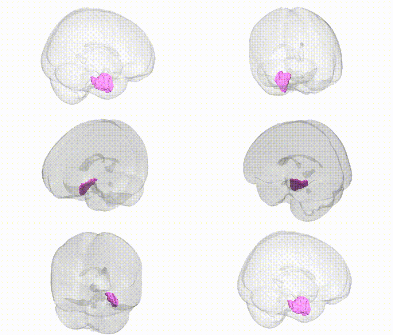
Full Quality Version: Download MP4
Wali Sidiqyar*, Gaurav Rudravaram*, Elyssa M. McMaster, Trent M. Schwartz, Adam M. Saunders, Kurt G. Schilling, Bennett A. Landman "Introducing SPINS: A Shared Public Visualization Library of Neuroanatomical Structures." Medical Imaging with Deep Learning- short paper
This resource is licensed under CC0 1.0 Universal (Public Domain).