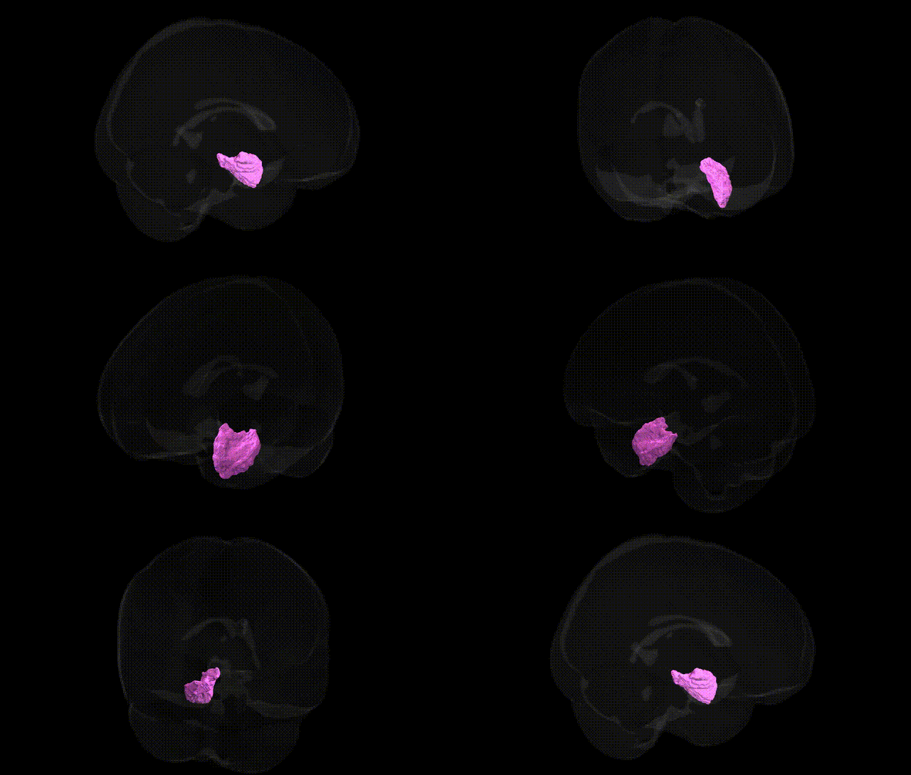
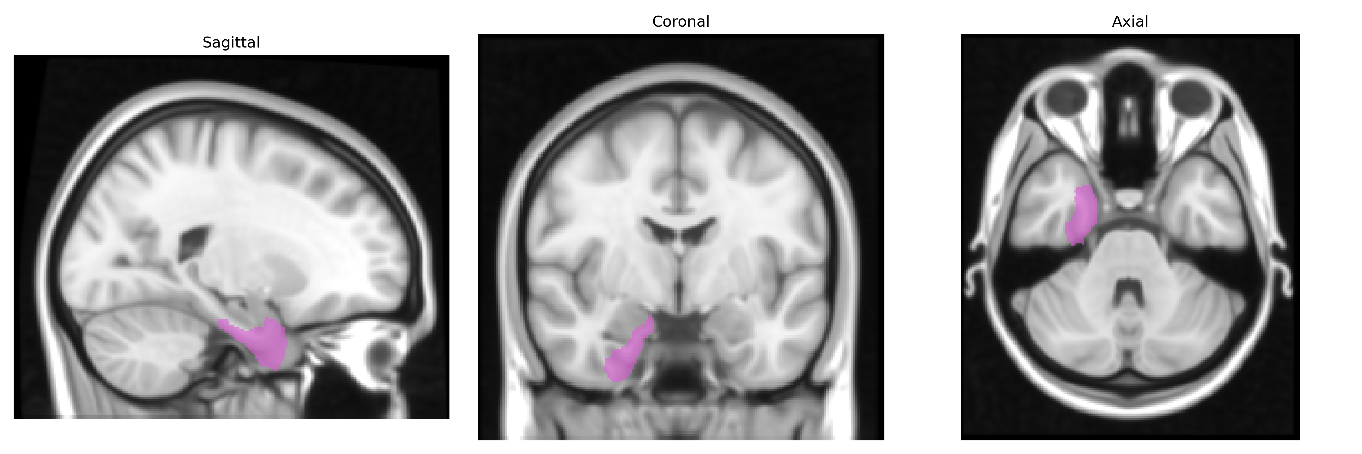
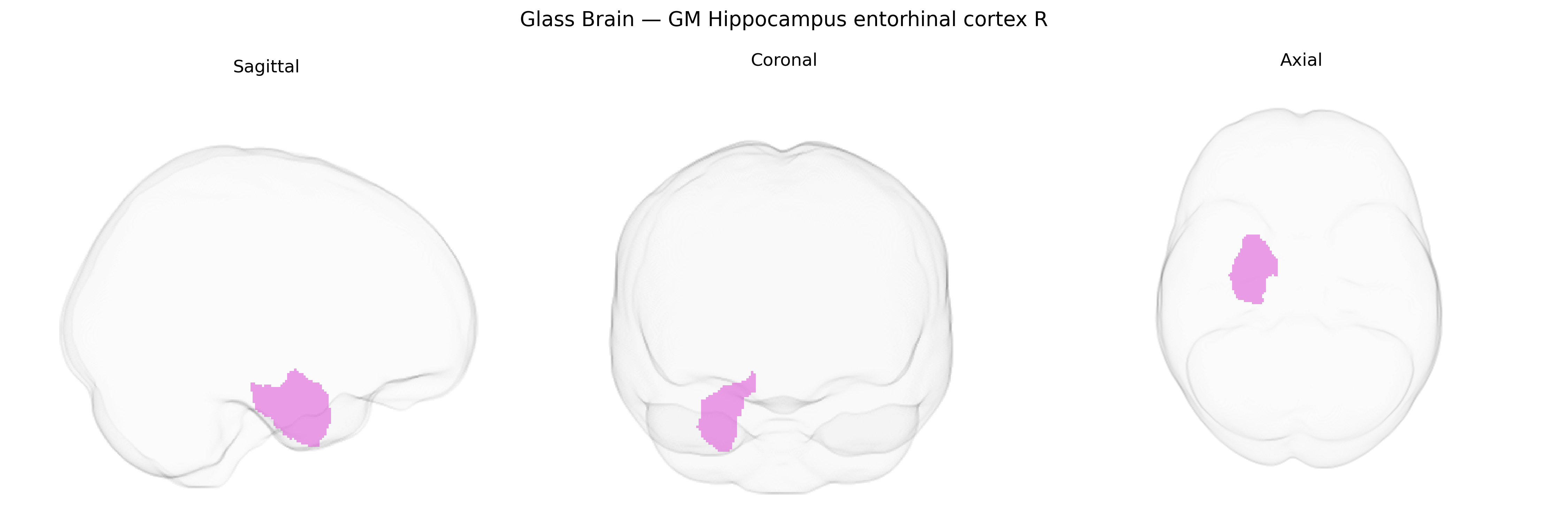
The bilateral GM Hippocampus entorhinal cortex R region in the Jülich maxprob thr25 1 mm atlas corresponds primarily to right entorhinal cortex and adjacent anterior hippocampal gray matter within the medial temporal lobe, forming a key hub of the parahippocampal-hippocampal memory network. This area receives highly processed multimodal cortical input and provides major projections to the hippocampus proper, supporting episodic memory formation, spatial navigation, and temporal-context coding. Cytoarchitectonically, it features a characteristic laminated structure distinct from neocortex and serves as a primary interface between neocortical association areas and hippocampal subfields such as dentate gyrus and CA regions. In humans, structural and functional changes in this region are strongly implicated in early Alzheimer’s disease and other neurodegenerative and memory disorders. No direct link: see Entorhinal cortex.
The right entorhinal cortex, part of the medial temporal lobe and continuous with the hippocampal formation, shows robust genetic associations with loci implicated in Alzheimer’s disease (AD), other dementias, and inter-individual variation in cortical structure. GWAS and imaging-genetics studies (e.g., ENIGMA, UK Biobank) have identified common variants in and near APOE (particularly the ε4-defining alleles in APOE and TOMM40) as major contributors to entorhinal cortical thinning, atrophy, and AD risk, with additional signals in genes involved in tau processing (MAPT on 17q21.31), immune and microglial function (TREM2, HLA region, CR1), and lipid metabolism and endocytosis (CLU, BIN1, PICALM). Polygenic risk for AD and related dementias strongly predicts reduced entorhinal cortical thickness and volume, and several large-scale GWAS of regional cortical morphology have reported significant SNP heritability for entorhinal structure, implicating loci in neuronal development, synaptic function, and neuroinflammation. The entorhinal cortex also emerges as a key region in genetic studies of age-related cognitive decline, mild cognitive impairment, and temporal lobe epilepsy, with risk variants in genes such as GRIN2A and SCN1A contributing to temporal lobe network susceptibility, and in psychiatric GWAS, polygenic risk for schizophrenia and major depression has modest but detectable associations with entorhinal and hippocampal volumetric variation.
Overview generated by GPT-4o (2026).
Region ID: 20
Hemisphere: bilateral
Atlas: Juelich maxprob thr25 1mm
Full Quality Version: Download MP4
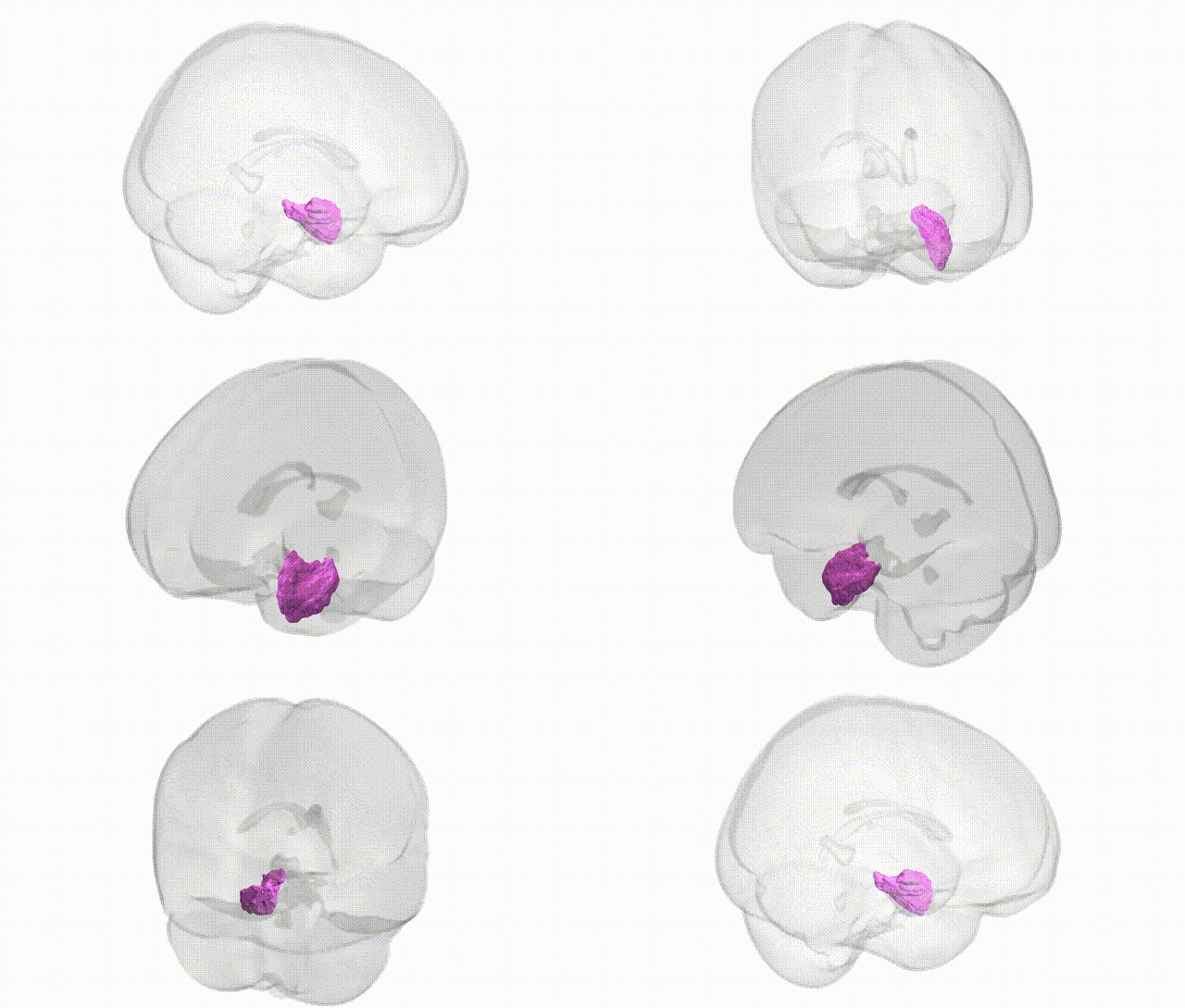
Full Quality Version: Download MP4
Wali Sidiqyar*, Gaurav Rudravaram*, Elyssa M. McMaster, Trent M. Schwartz, Adam M. Saunders, Kurt G. Schilling, Bennett A. Landman "Introducing SPINS: A Shared Public Visualization Library of Neuroanatomical Structures." Medical Imaging with Deep Learning- short paper
This resource is licensed under CC0 1.0 Universal (Public Domain).