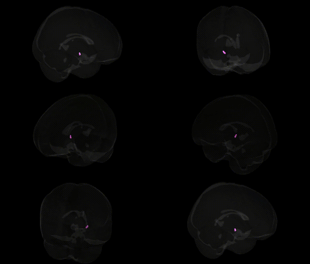
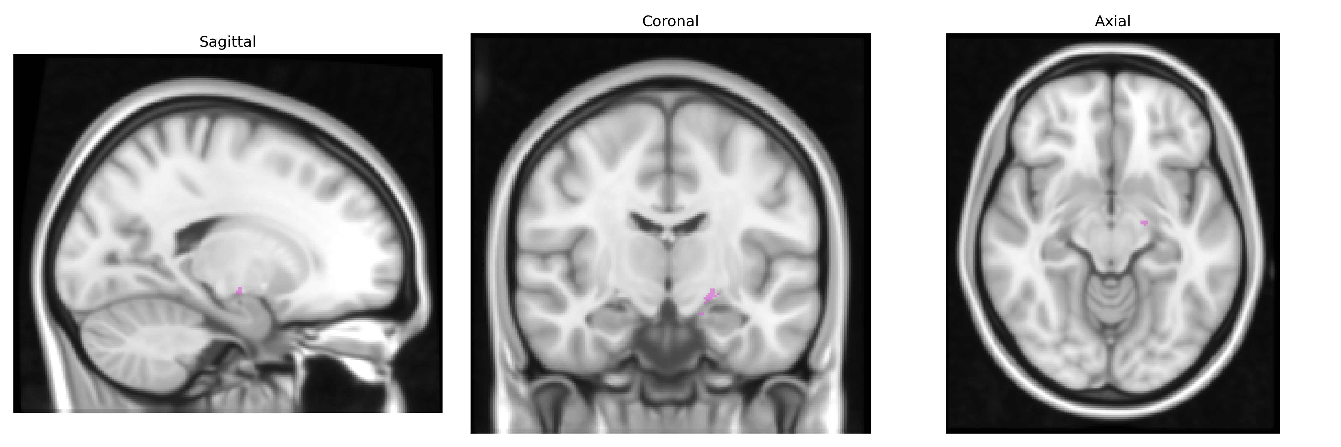
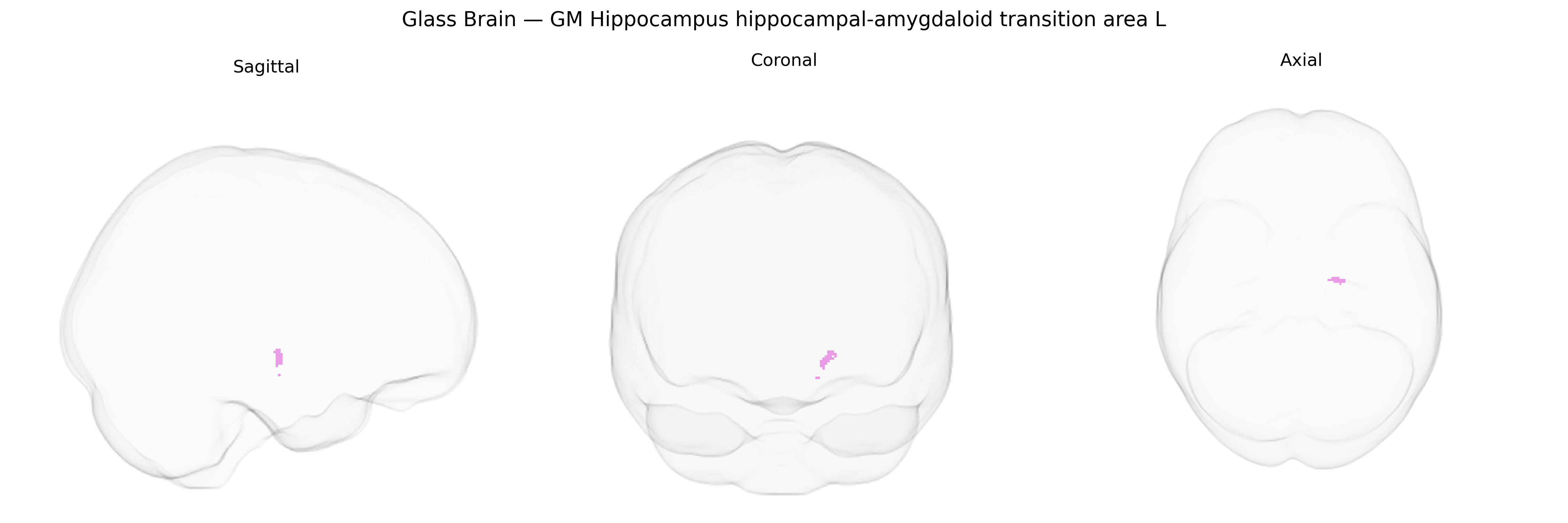
The bilateral GM Hippocampus hippocampal-amygdaloid transition area L, as defined in the Juelich maxprob thr25 1mm Atlas, refers to a gray matter region located at the anatomical interface between the hippocampus and the amygdala within the medial temporal lobe. This transition area participates in integrating mnemonic and emotional processes, acting as a conduit between hippocampal circuitry involved in declarative memory and spatial navigation and amygdalar networks that encode emotional salience and fear-related responses. Cytoarchitectonically, it exhibits features intermediate between hippocampal and amygdaloid tissue, reflecting its hybrid functional role in contextualizing emotionally relevant memories. There is no direct Wikipedia article for this specific transition area; a closely related structure is the Hippocampus.
The hippocampal–amygdaloid transition area (HATA) in the left hippocampus, as defined in the Juelich maxprob atlas, lies at the interface of hippocampal (e.g., cornu ammonis, subiculum) and amygdalar nuclei, and genetic associations reported for this region largely mirror those found for hippocampal and amygdalar volume and function more broadly. GWAS of hippocampal subfield volume and limbic structures implicate common variants in genes related to neurodevelopment, synaptic plasticity, and neurodegeneration (including loci in or near SORL1, MAPT, MSRB3, DPP4, and genes affecting glutamatergic signaling and axonal guidance), with some studies noting specific effects on anterior or medial hippocampal–amygdalar regions. Polygenic risk scores for Alzheimer’s disease, schizophrenia, and major depressive disorder show associations with reduced hippocampal and amygdalar volumes, and imaging–genetics work suggests that risk variants in APOE (particularly ε4), BDNF (Val66Met), COMT, CACNA1C, and several schizophrenia risk loci (e.g., in or near MIR137 and ZNF804A) modulate structure and connectivity in the medial temporal lobe, including the HATA region. Disorders linked via genetic and imaging–genetic evidence include Alzheimer’s disease and other dementias, temporal lobe epilepsy, schizophrenia and related psychoses, major depressive disorder, post‑traumatic stress disorder, and anxiety disorders, in which risk alleles and polygenic burdens are associated with altered HATA-adjacent hippocampal–amygdaloid morphology, microstructure, and functional coupling, although precise parcel-level GWAS specific to the Juelich HATA label remain sparse and findings are mostly inferred from broader hippocampal and amygdalar phenotypes.
Overview generated by GPT-4o (2026).
Region ID: 23
Hemisphere: bilateral
Atlas: Juelich maxprob thr25 1mm
Full Quality Version: Download MP4
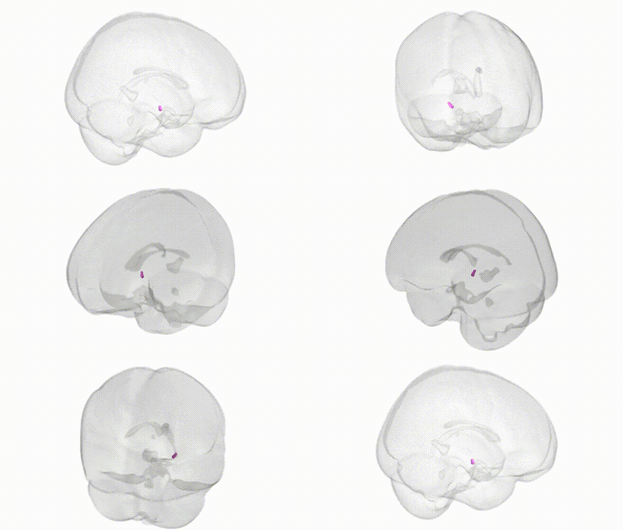
Full Quality Version: Download MP4
Wali Sidiqyar*, Gaurav Rudravaram*, Elyssa M. McMaster, Trent M. Schwartz, Adam M. Saunders, Kurt G. Schilling, Bennett A. Landman "Introducing SPINS: A Shared Public Visualization Library of Neuroanatomical Structures." Medical Imaging with Deep Learning- short paper
This resource is licensed under CC0 1.0 Universal (Public Domain).