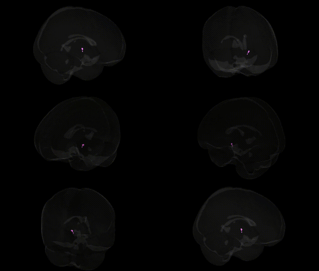
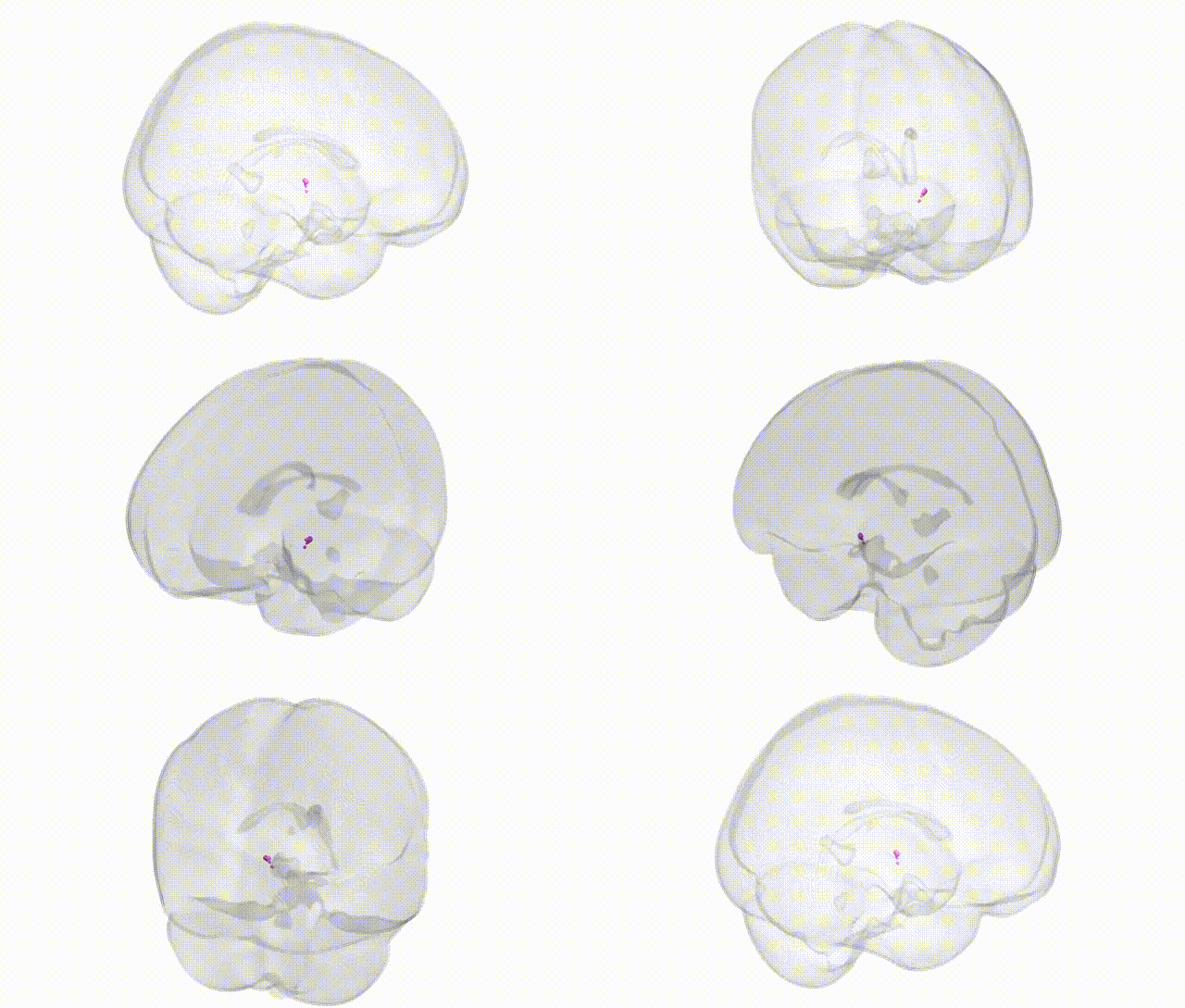
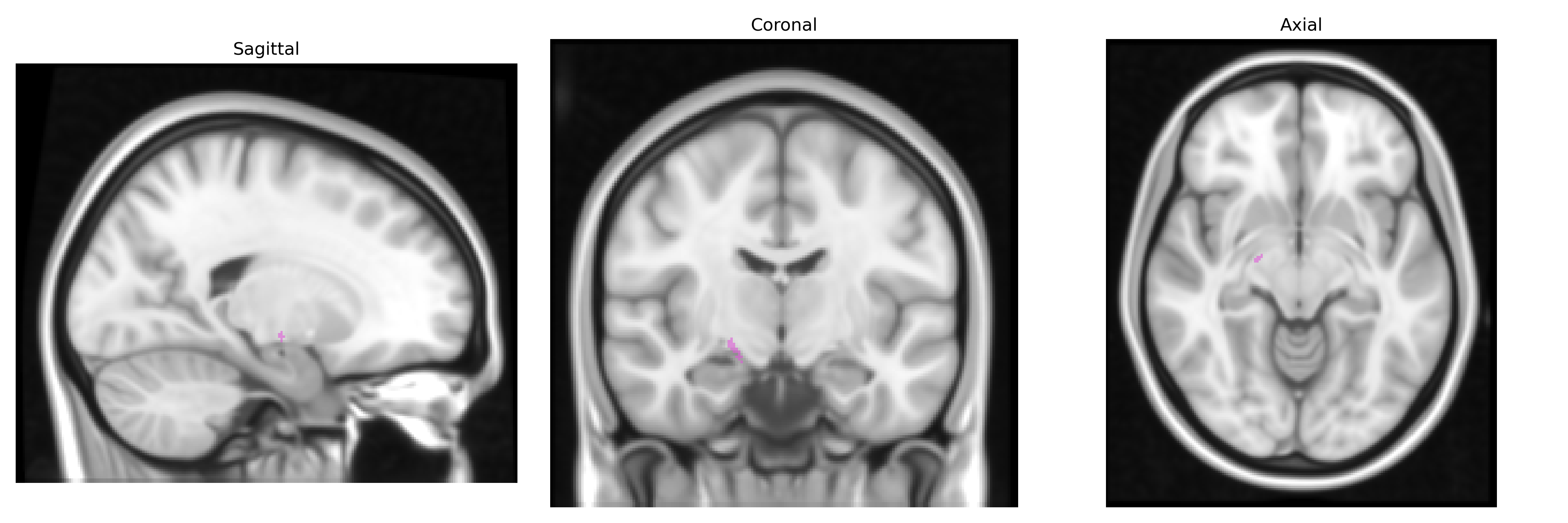
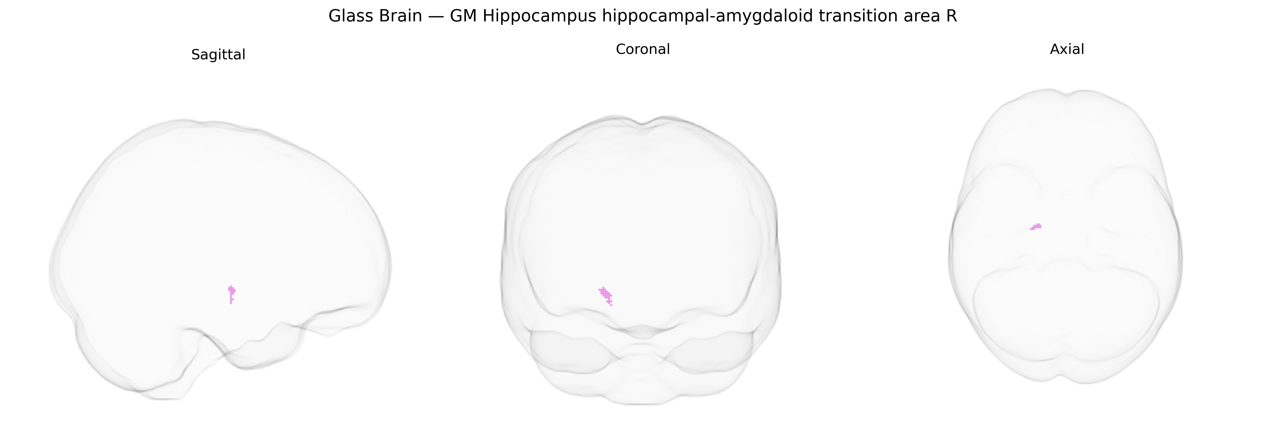
The bilateral GM Hippocampus hippocampal-amygdaloid transition area R, as defined in the Juelich maxprob thr25 1mm atlas, corresponds to a transitional zone between the hippocampal formation and the amygdala, located in the medial temporal lobe. This region encompasses cortex at the interface of the posterior amygdaloid complex and the anterior hippocampal head, including parts of the so-called hippocampal–amygdaloid transition area and adjacent entorhinal–perirhinal territory. It is implicated in integrating mnemonic and emotional information, serving as a conduit between hippocampal circuits involved in declarative memory and amygdalar circuits involved in affective processing and emotional salience. There is no direct link for this exact transition area; a related structure is the hippocampus: Hippocampus.
The hippocampal–amygdaloid transition area (HATA) is a medial temporal lobe interface region whose genetic associations are typically captured in GWAS of hippocampal or amygdalar subfield volumes and related limbic structures rather than as a separately labeled ROI, but several robust findings implicate genes influencing this region’s structure and function. Large-scale imaging–genetics studies (e.g., ENIGMA and UK Biobank) have identified common variants in genes such as MAPT, SLC6A4, APOE, BDNF, and KIBRA (WWC1), as well as loci in or near WWOX, MSRB3, and HRK, that associate with hippocampal volume, medial temporal lobe atrophy, or limbic connectivity, traits that anatomically encompass or closely border the HATA. These genetic signals are frequently linked to risk for Alzheimer’s disease (APOE, MAPT), major depressive disorder and anxiety (SLC6A4, BDNF), post-traumatic stress disorder (multiple loci affecting hippocampal volume and amygdala reactivity), schizophrenia and bipolar disorder (MAPT region and several polygenic risk loci affecting medial temporal structures), and epilepsy, especially mesial temporal lobe epilepsy with hippocampal sclerosis, where variants in immune, synaptic, and cell death pathways (e.g., HLA region, complement genes) have been associated with hippocampal pathology that extends into the hippocampal–amygdaloid transition. Additional GWAS of amygdala nuclei and hippocampal subfields report polygenic influences on this region through genes involved in neurodevelopment, synaptic plasticity, and stress reactivity, suggesting that variation in HATA structure and connectivity contributes to individual differences in emotional processing, memory, and vulnerability to neuropsychiatric and neurodegenerative disorders, even though the region is rarely isolated as a primary genetic target in current atlases and studies.
Overview generated by GPT-4o (2026).
Region ID: 24
Hemisphere: bilateral
Atlas: Juelich maxprob thr25 1mm
Full Quality Version: Download MP4
Full Quality Version: Download MP4
Wali Sidiqyar*, Gaurav Rudravaram*, Elyssa M. McMaster, Trent M. Schwartz, Adam M. Saunders, Kurt G. Schilling, Bennett A. Landman "Introducing SPINS: A Shared Public Visualization Library of Neuroanatomical Structures." Medical Imaging with Deep Learning- short paper
This resource is licensed under CC0 1.0 Universal (Public Domain).